Explore the intricate chambers, valves, and major blood vessels of the human heart with this detailed anatomical diagram, distinguishing pathways of oxygenated and deoxygenated blood. This article provides a comprehensive overview of cardiac structures, including the atria, ventricles, and key arteries and veins, essential for understanding cardiovascular function. Gain crucial insights into the heart’s sophisticated design and its role in maintaining life-sustaining circulation.
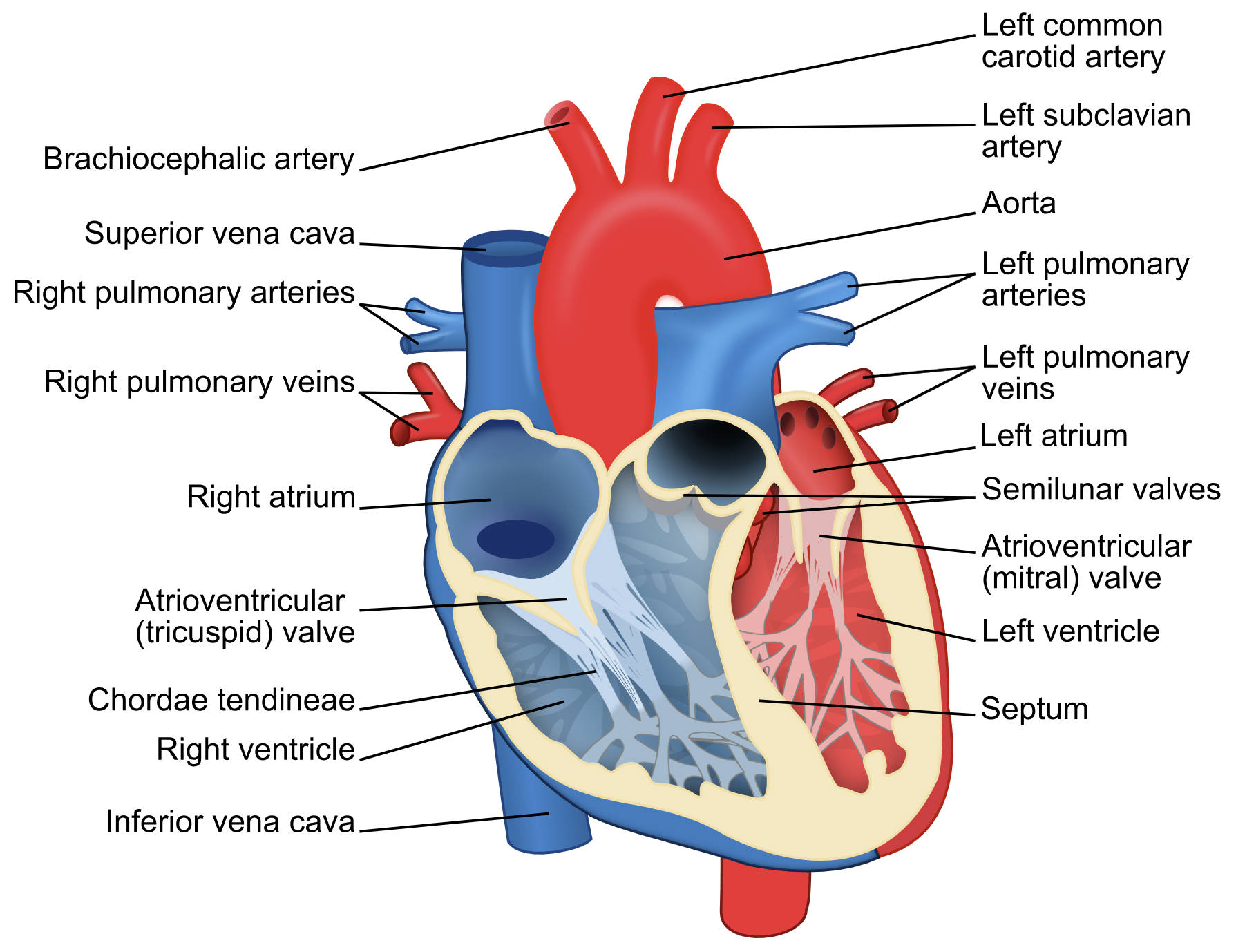
Brachiocephalic artery: This is the first and largest artery that branches off the aortic arch, located in the chest. It quickly divides into the right common carotid artery (supplying the head and neck) and the right subclavian artery (supplying the right arm and shoulder).
Superior vena cava: The superior vena cava is a large vein that carries deoxygenated blood from the upper half of the body (head, neck, upper limbs, and chest) to the right atrium of the heart. It is one of the two main vena cavae.
Right pulmonary arteries: These arteries carry deoxygenated blood from the right ventricle of the heart to the lungs. Unlike other arteries, pulmonary arteries transport deoxygenated blood, a critical part of the pulmonary circulation.
Right pulmonary veins: These veins carry oxygenated blood from the right lung back to the left atrium of the heart. Pulmonary veins are unique in that they carry oxygenated blood, in contrast to most other veins in the body.
Right atrium: The right atrium is the upper right chamber of the heart, which receives deoxygenated blood from the body via the superior and inferior vena cavae. It then pumps this blood through the tricuspid valve into the right ventricle.
Atrioventricular (tricuspid) valve: Located between the right atrium and the right ventricle, the tricuspid valve has three cusps. It prevents the backflow of blood into the right atrium during ventricular contraction (systole).
Chordae tendineae: These are fibrous cords that connect the cusps of the atrioventricular valves (tricuspid and mitral valves) to the papillary muscles in the ventricles. They prevent the valve cusps from inverting into the atria during ventricular contraction.
Right ventricle: The right ventricle is the lower right chamber of the heart, responsible for pumping deoxygenated blood into the pulmonary trunk, which then leads to the lungs for oxygenation. Its muscular walls are thinner than the left ventricle’s, reflecting the lower pressure required for pulmonary circulation.
Inferior vena cava: The inferior vena cava is the largest vein in the body, carrying deoxygenated blood from the lower half of the body (legs, abdomen, and pelvis) to the right atrium of the heart. It joins the superior vena cava in delivering blood for pulmonary circulation.
Left common carotid artery: This major artery branches directly from the aortic arch and ascends into the neck, supplying oxygenated blood to the left side of the head and neck, including the brain. It is crucial for cerebral circulation.
Left subclavian artery: Also branching directly from the aortic arch, the left subclavian artery supplies oxygenated blood to the left arm, shoulder, and part of the chest. It gives rise to several branches, including the vertebral artery.
Aorta: The aorta is the largest artery in the body, originating from the left ventricle and arching over the heart before descending. It distributes oxygenated blood from the heart to all systemic arteries, maintaining the body’s circulation.
Left pulmonary arteries: These arteries carry deoxygenated blood from the right ventricle to the left lung. Like their right counterparts, they are part of the pulmonary circulation system.
Left pulmonary veins: These veins carry oxygenated blood from the left lung back to the left atrium of the heart. They complete the pulmonary circuit, bringing oxygen-rich blood for systemic distribution.
Left atrium: The left atrium is the upper left chamber of the heart, receiving oxygenated blood from the lungs via the pulmonary veins. It then pumps this oxygenated blood through the mitral valve into the left ventricle.
Semilunar valves: These valves, which include the aortic and pulmonary valves, are located at the exits of the ventricles (where the aorta and pulmonary trunk begin). They prevent the backflow of blood into the ventricles during ventricular relaxation (diastole) and have a crescent moon shape.
Atrioventricular (mitral) valve: Also known as the bicuspid valve, this valve is situated between the left atrium and the left ventricle and has two cusps. It ensures that oxygenated blood flows only from the left atrium into the left ventricle, preventing regurgitation.
Left ventricle: The left ventricle is the strongest and most muscular chamber of the heart, responsible for pumping oxygenated blood into the aorta to be distributed throughout the entire body. Its thick walls generate the high pressure necessary for systemic circulation.
Septum: The septum is the muscular wall that divides the heart into right and left sides, preventing the mixing of deoxygenated blood (on the right side) with oxygenated blood (on the left side). This complete separation is vital for the efficiency of the double circulatory system.
The human heart is a complex, four-chambered muscular organ that serves as the central pump of the cardiovascular system. Its primary function is to circulate blood throughout the body, delivering vital oxygen and nutrients to tissues while removing metabolic waste products. This remarkable feat is achieved through a precisely coordinated sequence of contractions and relaxations, driven by an intricate electrical system. A detailed understanding of the heart’s anatomy, including its chambers, valves, and associated great vessels, is fundamental to comprehending its physiological functions and the pathophysiology of cardiac diseases. The provided diagram offers a comprehensive view, distinguishing pathways of oxygenated (red) and deoxygenated (blue) blood.
The heart operates as a double pump, facilitating two distinct circulatory loops: the pulmonary circulation and the systemic circulation. In pulmonary circulation, deoxygenated blood from the body enters the right atrium, passes into the right ventricle, and is then pumped to the lungs via the pulmonary arteries. In the lungs, blood releases carbon dioxide and picks up oxygen. This newly oxygenated blood then returns to the left atrium via the pulmonary veins. The systemic circulation begins as this oxygenated blood moves from the left atrium into the left ventricle, which then powerfully pumps it into the aorta for distribution to the rest of the body.
The unidirectional flow of blood through the heart is maintained by four crucial valves: the two atrioventricular (AV) valves (tricuspid and mitral) and the two semilunar valves (pulmonary and aortic). The AV valves prevent backflow into the atria during ventricular contraction, while the semilunar valves prevent backflow into the ventricles during relaxation. The muscular septum effectively divides the heart, ensuring complete separation of oxygenated and deoxygenated blood, which is critical for maintaining high metabolic rates in mammals. This intricate design underscores the heart’s efficiency in supporting all bodily functions.
Key anatomical structures and their roles include:
- Chambers: Atria (receiving), Ventricles (pumping).
- Valves: Regulate unidirectional blood flow.
- Major Arteries: Aorta (systemic), Pulmonary Trunk (pulmonary).
- Major Veins: Vena Cavae (systemic), Pulmonary Veins (pulmonary).
- Septum: Divides oxygenated and deoxygenated blood.
In conclusion, the human heart is a masterpiece of biological engineering, with its precisely coordinated chambers, valves, and major blood vessels orchestrating a continuous, life-sustaining flow of blood. A thorough understanding of each component, from the aorta distributing oxygenated blood to the body to the right ventricle pumping deoxygenated blood to the lungs, is indispensable for grasping the complexities of cardiovascular physiology. This anatomical knowledge is paramount for healthcare professionals in diagnosing, treating, and managing the myriad of cardiac conditions that can affect this vital organ, ultimately contributing to improved patient outcomes and overall public health.

