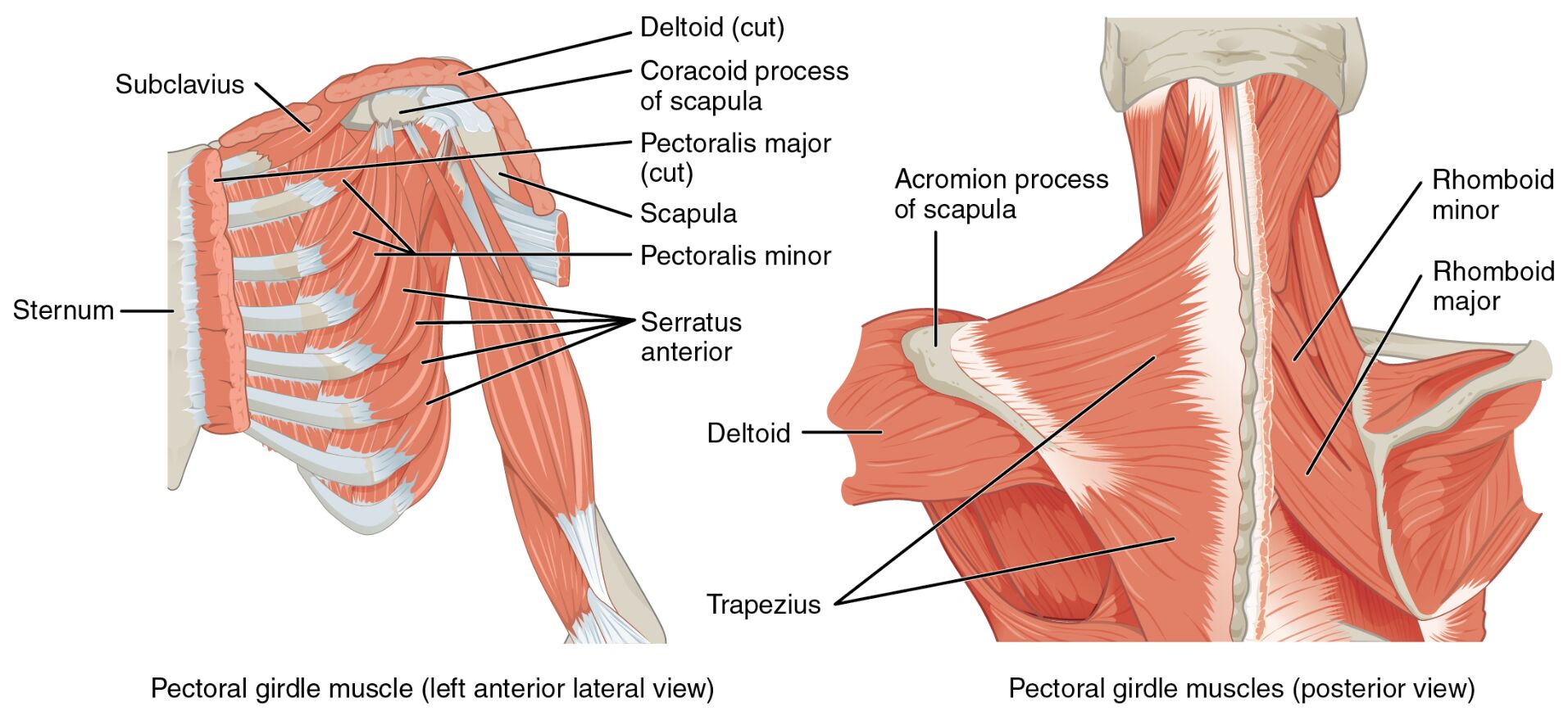
The muscles that position the pectoral girdle are essential for providing a stable base that enables arm movement, working beneath the surface to support the shoulder complex. This detailed exploration of the muscles that position the pectoral girdle reveals their deeper anatomy, with the pectoralis major and deltoid cut away to highlight their roles, offering valuable insights into upper body mechanics.
Labeled Parts Introduction
Clavicle
The clavicle is a slender S-shaped bone connecting the sternum to the scapula, serving as a key attachment point for muscles that stabilize the pectoral girdle, and provides leverage for arm movements. It supports the weight of the upper limb and maintains the shoulder’s position.
Scapula
The scapula is a flat, triangular bone on the upper back, acting as the primary attachment site for muscles that position the pectoral girdle, and facilitates a wide range of shoulder motions. It moves dynamically with muscle contractions to adjust the arm’s range of motion.
Serratus anterior
The serratus anterior is a fan-shaped muscle along the lateral chest, originating from the upper ribs and inserting into the scapula, and protracts the scapula while stabilizing the pectoral girdle. It prevents winging of the scapula and supports pushing movements.
Pectoralis minor
The pectoralis minor is a thin muscle beneath the pectoralis major, attaching from the ribs to the scapula, and depresses the scapula while aiding in pectoral girdle stability. It also assists in elevating the ribs during deep inspiration.
Subclavius
The subclavius is a small muscle located beneath the clavicle, connecting it to the first rib, and stabilizes the clavicle while protecting underlying neurovascular structures. It helps prevent excessive upward movement of the clavicle during arm elevation.
Trapezius
The trapezius is a large, diamond-shaped muscle covering the upper back and neck, attaching to the clavicle and scapula, and elevates, retracts, and rotates the scapula to position the pectoral girdle. It plays a crucial role in posture and upper limb movement.
Levator scapulae
The levator scapulae is a slender muscle running from the cervical vertebrae to the scapula, elevating the scapula and assisting in neck extension, and contributes to pectoral girdle stability. It helps maintain shoulder height during arm raising.
Rhomboid major
The rhomboid major is a broad muscle beneath the trapezius, connecting the spinal column to the scapula, and retracts and stabilizes the scapula to support the pectoral girdle. It works with the rhomboid minor to hold the scapula against the thoracic wall.
Rhomboid minor
The rhomboid minor is a smaller muscle above the rhomboid major, linking the cervical and thoracic vertebrae to the scapula, and retracts the scapula while aiding in its stabilization. It enhances the coordinated movement of the pectoral girdle.
Overview of Pectoral Girdle Muscle Anatomy
The muscles that position the pectoral girdle provide a stable foundation for arm movement, with the clavicle and scapula serving as critical anchoring points. This image, with the pectoralis major and deltoid cut away, exposes the deeper serratus anterior, trapezius, and other muscles that ensure shoulder stability. Their intricate interplay is fundamental to upper body function.
- Creates a dynamic base that supports the humerus during lifting or pushing.
- Protects the shoulder joint by maintaining proper alignment.
Structure of Pectoral Girdle Positioning Muscles
The trapezius and levator scapulae extend from the spine to the scapula, while the rhomboid major and rhomboid minor reinforce scapular retraction. The pectoralis minor and subclavius attach to the clavicle, stabilizing it, with the serratus anterior anchoring the scapula to the ribs.
- The trapezius covers a wide area, enabling diverse scapular movements.
- The serratus anterior provides lateral support to the pectoral girdle.
Role in Shoulder Stability and Movement
The serratus anterior protracts the scapula during pushing actions, while the trapezius and rhomboid major retract it for pulling motions. The levator scapulae and pectoralis minor adjust scapular height and depression, ensuring the clavicle remains stable via the subclavius.
- The rhomboid minor enhances scapular adduction for posture.
- The subclavius minimizes clavicular stress during arm elevation.
Clinical Relevance and Physical Health
The serratus anterior is often weakened in scapular winging, a condition requiring targeted strengthening. The trapezius and levator scapulae may become tight, contributing to neck pain, while the pectoralis minor tightness can affect shoulder posture.
- A strained rhomboid major can limit scapular retraction.
- The clavicle serves as a diagnostic point for pectoral girdle injuries.
Physical Examination and Rehabilitation
Physical exams assess the scapula for movement and the clavicle for alignment, with the trapezius checked for tension. Rehabilitation focuses on strengthening the serratus anterior and stretching the levator scapulae to restore shoulder function.
- Proper scapula positioning enhances upper limb mobility.
- The pectoralis minor is targeted to relieve shoulder impingement.
Conclusion
The muscles that position the pectoral girdle, as shown with the pectoralis major and deltoid dissected, form a robust system that stabilizes the clavicle and scapula for arm movement. From the serratus anterior to the rhomboid minor, these muscles ensure shoulder integrity and function. Understanding their anatomy supports effective diagnosis and treatment of upper body conditions.

