The muscles that move the humerus are essential for a wide range of arm movements, originating from various points across the body to facilitate flexion, abduction, and rotation. This detailed exploration of the muscles that move the humerus, as depicted in this anatomical image, highlights their diverse origins and insertions, providing valuable insights into upper limb function.
Labeled Parts Introduction
Clavicle
The clavicle is a slender, S-shaped bone connecting the sternum to the scapula, serving as an attachment point for muscles that influence humerus movement, and provides leverage for arm actions. It supports the deltoid and other muscles during abduction.
Scapula
The scapula is a flat, triangular bone on the back, acting as the origin for multiple muscles that move the humerus, and facilitates a broad range of shoulder motions. It serves as a stable base for muscles like the subscapularis and infraspinatus.
Sternum
The sternum is the flat bone at the front of the chest, providing an origin for the pectoralis major, and contributes to the structural framework of the thoracic cage. It supports the anterior muscles involved in humerus flexion.
Humerus
The humerus is the long bone of the upper arm, serving as the insertion point for muscles that move it, and is central to arm flexion, abduction, and rotation. Its articulation with the scapula allows for a wide range of motions driven by these muscles.
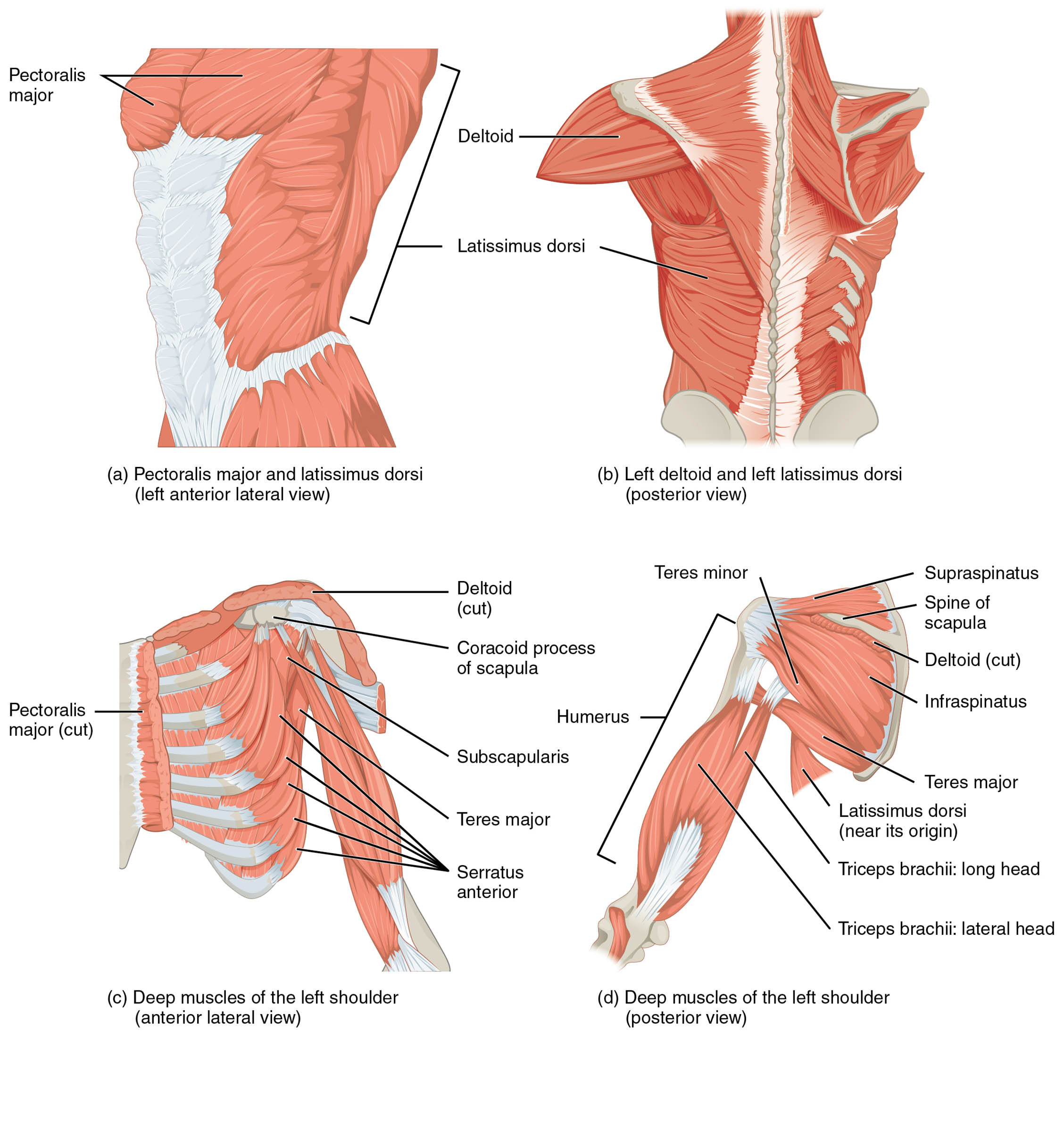
Pectoralis major
The pectoralis major is a large, fan-shaped muscle on the anterior chest, originating from the sternum and clavicle, and flexes, adducts, and internally rotates the humerus. It plays a key role in pushing movements and stabilizing the shoulder joint.
Deltoid
The deltoid is a thick, triangular muscle covering the shoulder, originating from the clavicle and scapula, and abducts, flexes, and extends the humerus depending on the portion activated. It is crucial for lifting the arm and providing shoulder contour.
Subscapularis
The subscapularis is a deep muscle on the anterior scapula, originating from its inner surface, and internally rotates the humerus while stabilizing the shoulder joint. It is a primary component of the rotator cuff, protecting the glenohumeral joint.
Latissimus dorsi
The latissimus dorsi is a broad muscle of the lower and middle back, originating from the spine and iliac crest, and extends, adducts, and internally rotates the humerus. It is powerful in pulling movements, such as rowing or climbing.
Infraspinatus
The infraspinatus is a posterior muscle below the scapular spine, originating from the scapula, and externally rotates the humerus while stabilizing the shoulder. It is another vital rotator cuff muscle, aiding in arm positioning.
Teres major
The teres major is a thick muscle on the posterior scapula, originating near the inferior angle, and adducts and internally rotates the humerus while assisting in extension. It works closely with the latissimus dorsi to support arm movement.
Teres minor
The teres minor is a narrow muscle below the infraspinatus, originating from the scapula, and externally rotates the humerus while contributing to shoulder stability. It is part of the rotator cuff, enhancing joint integrity during motion.
Supraspinatus
The supraspinatus is a small muscle above the scapular spine, originating from the supraspinous fossa, and initiates abduction of the humerus while stabilizing the shoulder joint. It is a key rotator cuff muscle involved in the early phase of arm lifting.
Overview of Humerus Moving Muscle Anatomy
The muscles that move the humerus are strategically positioned to enable a variety of arm motions, with origins ranging from the sternum to the scapula and lower back. This image illustrates the pectoralis major, deltoid, and latissimus dorsi, among others, with their roles in anterior, superior, and posterior movements clearly defined. Their coordinated action is fundamental to upper limb functionality.
- Supports complex movements like lifting, pushing, and pulling.
- Maintains shoulder joint stability during dynamic activities.
Structure of Humerus Moving Muscles
The pectoralis major and subscapularis originate anteriorly, with the deltoid spanning the shoulder from the clavicle and scapula. The latissimus dorsi arises from the lower back, while the infraspinatus, teres major, teres minor, and supraspinatus attach to the posterior and superior scapula, forming a robust muscular network.
- The deltoid is divided into three parts for versatile arm action.
- The subscapularis lies deep, protecting the shoulder’s anterior aspect.
Role in Arm Movement
The pectoralis major flexes and adducts the humerus anteriorly, while the deltoid abducts it superiorly from its scapular and clavicular origins. The latissimus dorsi and teres major move the humerus inferiorly and posteriorly, with the infraspinatus, teres minor, and supraspinatus fine-tuning rotation and stability.
- The supraspinatus is critical for the initial lift in arm abduction.
- The infraspinatus and teres minor ensure smooth external rotation.
Clinical Relevance and Physical Health
The rotator cuff muscles, including the supraspinatus and infraspinatus, are prone to tears, leading to shoulder pain and weakness. The latissimus dorsi may be strained during heavy lifting, affecting posterior humerus movement, while deltoid imbalances can cause shoulder instability.
- A torn subscapularis can impair internal rotation and joint protection.
- The pectoralis major is often assessed for chest tightness or injury.
Physical Examination and Rehabilitation
Physical exams evaluate the deltoid for abduction strength and the latissimus dorsi for extension power, with the rotator cuff muscles checked for range of motion. Rehabilitation focuses on strengthening the teres minor and stretching the pectoralis major to restore shoulder function.
- Proper supraspinatus function supports early abduction phases.
- The teres major is targeted to enhance posterior arm strength.
Conclusion
The muscles that move the humerus, as shown in this anatomical view, form a sophisticated system that drives arm flexion, abduction, and rotation from multiple origins. From the pectoralis major to the supraspinatus, these muscles ensure precise movement and shoulder stability. Understanding their anatomy provides a solid foundation for diagnosing and treating upper limb conditions.

