The lower leg is a dynamic region supported by a sophisticated network of muscles that enable a wide range of movements. This article delves into the muscles of the lower leg, presented through detailed diagrams of the right leg in anterior, superficial posterior, and deep posterior views, highlighting their anatomical structure and functional roles. These muscles, categorized into anterior and posterior compartments, are primarily responsible for dorsiflexion and plantar flexion, respectively, while lateral and medial muscles assist in inverting, everting, and rotating the foot. By examining the labeled illustrations, readers can gain a comprehensive understanding of these muscles’ contributions to leg and foot function.
Introduction to the Lower Leg Muscles
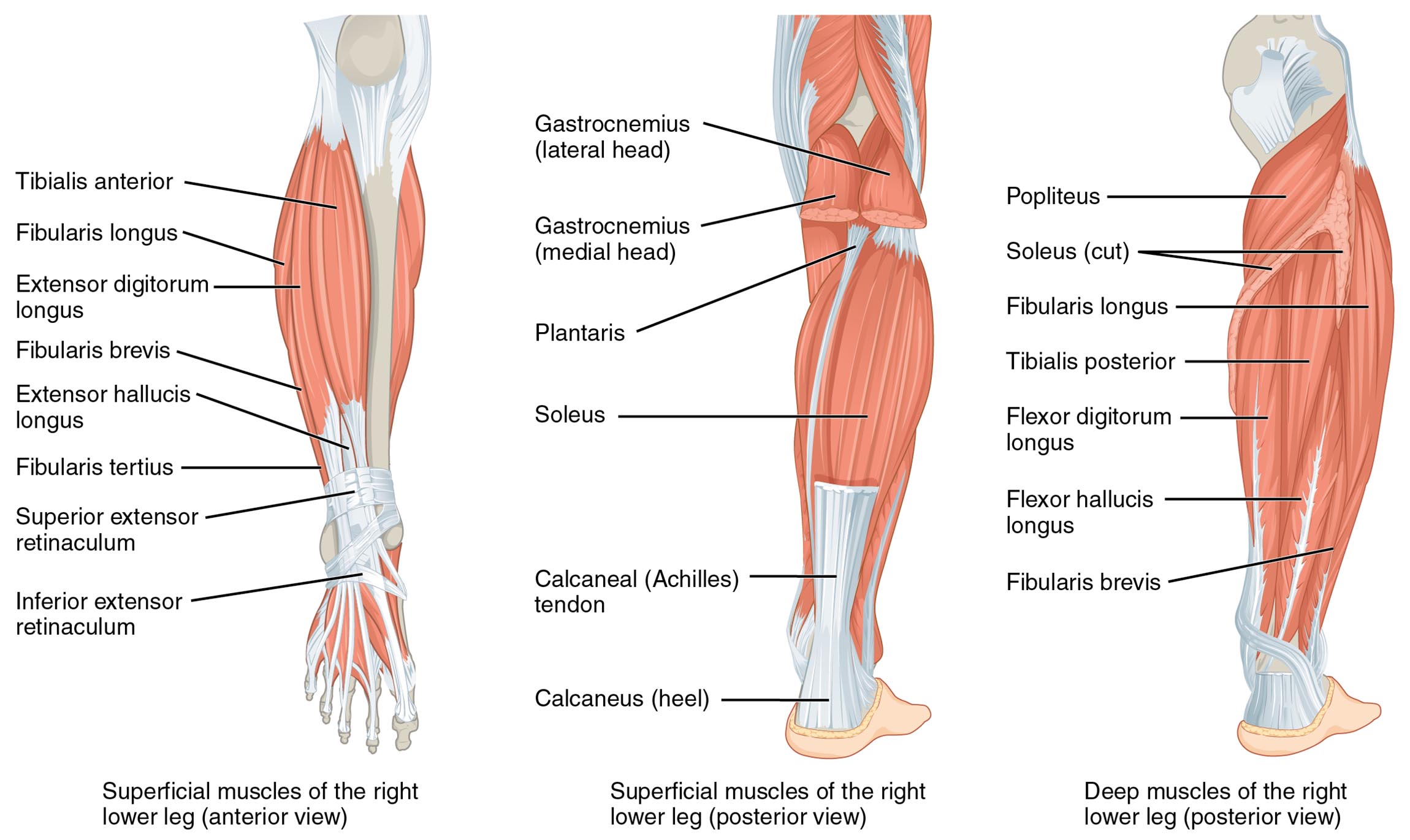
The muscles of the lower leg form a critical component of the lower limb’s muscular system. Their organization across different views reveals their specialized roles in movement. This section reviews the labeled structures that define their anatomy and function.
- Tibialis anterior: Located on the anterior compartment, this muscle dorsiflexes and inverts the foot. It plays a key role in lifting the foot during walking.
- Fibularis longus: Positioned on the lateral side, this muscle everts and plantar flexes the foot. It supports stability on uneven surfaces.
- Extensor digitorum longus: Found in the anterior compartment, it extends the toes and dorsiflexes the foot. It aids in toe lifting during gait.
- Fibularis brevis: Located laterally, this muscle everts the foot and assists in plantar flexion. It enhances lateral ankle stability.
- Extensor hallucis longus: Positioned in the anterior compartment, it extends the big toe and dorsiflexes the foot. It is essential for precise toe movements.
- Fibularis tertius: Found on the anterior lateral side, it dorsiflexes and everts the foot. It assists in foot elevation during motion.
- Superior extensor retinaculum: A band on the anterior ankle, it holds extensor tendons in place. It ensures smooth tendon movement during dorsiflexion.
- Inferior extensor retinaculum: Another anterior band, it stabilizes extensor tendons. It prevents tendon displacement during foot motion.
- Gastrocnemius (lateral head): Part of the posterior compartment, it plantar flexes the foot and flexes the knee. It contributes to the power of the calf.
- Gastrocnemius (medial head): Also in the posterior compartment, it plantar flexes the foot and flexes the knee. It works with the lateral head for calf strength.
- Plantaris: A small posterior muscle, it weakly plantar flexes the foot. It assists in knee flexion and provides tendon support.
- Soleus: Located deep in the posterior compartment, it plantar flexes the foot. It is vital for standing and maintaining posture.
- Calcaneal (Achilles) tendon: Connects the calf muscles to the heel, it transmits force for plantar flexion. It is crucial for walking and jumping.
- Calcaneus (heel): The heel bone serves as an insertion point for the Achilles tendon. It anchors the posterior muscles for stability.
- Popliteus: A deep posterior muscle, it flexes and rotates the knee internally. It unlocks the knee joint during movement.
- Fibularis longus: Repeated in the posterior view, it everts and plantar flexes the foot. It supports lateral ankle stability from behind.
- Tibialis posterior: A deep posterior muscle, it inverts and plantar flexes the foot. It maintains the medial arch of the foot.
- Flexor digitorum longus: Located deep posteriorly, it flexes the toes and plantar flexes the foot. It aids in gripping the ground.
- Flexor hallucis longus: A deep posterior muscle, it flexes the big toe and plantar flexes the foot. It supports toe stability during push-off.
- Fibularis brevis: Repeated in the posterior view, it everts the foot and assists in plantar flexion. It enhances lateral ankle support.
The muscles of the lower leg‘s diverse placement enables complex movements. Their labeled views provide a clear picture of their anatomical and functional roles.
Functional Roles of the Lower Leg Muscles
The muscles of the lower leg are essential for a variety of movements. Their compartmental organization supports specific actions across the leg and foot. This section outlines their functional contributions.
- The tibialis anterior and extensor hallucis longus dorsiflex the foot. These actions lift the foot, preventing tripping during walking.
- The fibularis longus and fibularis brevis evert the foot. They stabilize the ankle on uneven terrain, aiding in balance.
- The gastrocnemius and soleus plantar flex the foot. This movement powers pushing off during walking or running.
- The tibialis posterior and flexor digitorum longus invert the foot. They maintain the medial arch, supporting foot structure.
- The popliteus internally rotates the knee. This action unlocks the knee, facilitating smooth leg bending.
- The calcaneal tendon transmits force for plantar flexion. It ensures efficient energy transfer from the calf to the heel.
The muscles of the lower leg‘s coordinated actions enhance mobility. Their specific roles support both stability and dynamic movement.
Clinical Significance and Practical Applications
The muscles of the lower leg are frequently assessed in clinical evaluations of leg function. Their condition directly impacts mobility and daily activities. This section explores their clinical importance.
- Strain in the gastrocnemius can cause calf pain or Achilles tendon issues. Stretching and strengthening exercises help restore function.
- Weakness in the tibialis anterior may lead to foot drop. Targeted therapy improves dorsiflexion and prevents tripping.
- Injury to the fibularis longus can impair eversion, affecting ankle stability. Rehabilitation focuses on rebuilding lateral support.
- Tightness in the soleus may contribute to plantar fasciitis. Stretching alleviates heel pain and enhances flexibility.
- Understanding their anatomy aids in diagnosing conditions like posterior tibial tendon dysfunction. This knowledge guides effective treatment plans.
This insight is valuable for professionals addressing leg issues. The muscles of the lower leg‘s roles underscore the need for precise therapeutic interventions.
Conclusion
The muscles of the lower leg, as illustrated in the anterior, superficial posterior, and deep posterior views of the right leg, showcase the intricate design of the lower limb. This article has explored their anatomical structure, diverse functional roles, and clinical significance, providing a thorough understanding of their importance. From the tibialis anterior enabling dorsiflexion to the gastrocnemius powering plantar flexion, each muscle contributes uniquely to leg and foot mobility. Continued study of these muscles will enhance therapeutic strategies and deepen appreciation for the complex mechanics of lower leg movement.

