The accessory nerve, a key cranial nerve, plays a vital role in coordinating movements of the head, neck, and shoulders by innervating the sternocleidomastoid and trapezius muscles. This article explores an image detailing these muscles, their attachments, and their synergistic and antagonistic actions, providing a comprehensive understanding of their anatomical and functional significance.
Accessory nerve The accessory nerve, or cranial nerve XI, originates from the brainstem and spinal cord, providing motor innervation to the sternocleidomastoid and trapezius muscles. It enables head rotation, neck flexion, and shoulder elevation through its nerve fibers.
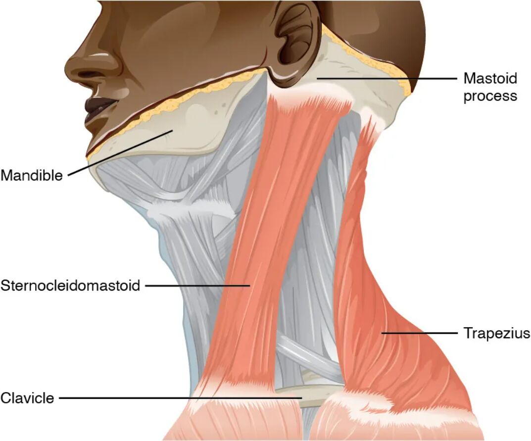
Sternocleidomastoid The sternocleidomastoid muscle runs from the sternum and clavicle to the mastoid process, allowing head rotation and flexion. It works with or against the trapezius depending on the movement, contributing to neck mobility.
Trapezius The trapezius muscle extends from the occipital bone and cervical/thoracic vertebrae to the clavicle and scapula, facilitating shoulder elevation and head extension. It acts as a synergist with the sternocleidomastoid in lateral flexion or as an antagonist in head movements.
Mastoid process The mastoid process is a bony projection behind the ear, serving as an attachment point for the sternocleidomastoid. It anchors the muscle, enabling effective head rotation and tilting.
Sternum The sternum, or breastbone, provides a lower attachment for the sternocleidomastoid, stabilizing the muscle’s origin. It supports movements involving neck flexion and rotation.
Clavicle The clavicle, or collarbone, serves as an attachment site for both the sternocleidomastoid and trapezius, aiding in head and shoulder movements. It acts as a structural link between the arm and torso.
Scapula The scapula, or shoulder blade, is a key attachment for the trapezius, supporting shoulder elevation and retraction. It enhances upper body stability and mobility.
Occipital bone The occipital bone, at the base of the skull, anchors the trapezius’ upper fibers, facilitating head extension. It provides a stable base for neck and shoulder coordination.
Cervical vertebrae The cervical vertebrae, part of the neck’s spinal column, attach the trapezius and support its role in head and shoulder movements. They allow flexibility and strength in the upper spine.
Thoracic vertebrae The thoracic vertebrae, in the upper back, provide additional attachment points for the trapezius, enhancing its role in shoulder elevation. They contribute to the muscle’s broad range of motion.
Anatomical Overview of Accessory Nerve-Controlled Muscles
The accessory nerve innervates two major muscles with distinct attachments. This innervation supports a range of head and shoulder movements.
- The accessory nerve originates from the brainstem and upper spinal cord.
- The sternocleidomastoid connects the sternum, clavicle, and mastoid process.
- The trapezius spans from the occipital bone to the scapula via cervical and thoracic vertebrae.
- These muscles work together or oppositely based on movement demands.
- Their attachments ensure coordinated upper body function.
Functional Roles in Head and Shoulder Movement
The sternocleidomastoid and trapezius perform complementary and opposing actions. This duality enhances neck and shoulder mobility.
- The sternocleidomastoid rotates the head when acting unilaterally.
- It flexes the neck when both sides contract together.
- The trapezius elevates the shoulders and extends the head.
- Both muscles act as synergists in lateral flexion toward the shoulder.
- Their antagonistic action balances head flexion and extension.
Attachment Points and Structural Support
The muscles’ attachment sites provide stability and leverage for movement. These points are critical for effective muscle function.
- The mastoid process anchors the sternocleidomastoid for head rotation.
- The sternum and clavicle offer a solid base for neck flexion.
- The scapula and clavicle support trapezius-mediated shoulder elevation.
- The occipital bone and cervical/thoracic vertebrae enhance head extension.
- This network ensures robust support for upper body dynamics.
Clinical Relevance and Related Conditions
Understanding these muscles aids in diagnosing and treating nerve or muscle disorders. This knowledge supports targeted interventions.
- Accessory nerve injury can cause weakness in the sternocleidomastoid and trapezius.
- Torticollis, or wry neck, may result from sternocleidomastoid dysfunction.
- Shoulder droop or scapular winging indicates trapezius impairment.
- Physical therapy or surgery may address nerve damage effects.
- Electromyography assesses muscle and nerve health.
The accessory nerve’s control over the sternocleidomastoid and trapezius muscles, with their extensive attachments from the mastoid process to the scapula, enables a wide range of head and shoulder movements. Their synergistic and antagonistic actions support posture and mobility, while conditions like torticollis highlight the importance of this system. This detailed exploration provides valuable insights into muscular anatomy and informs clinical approaches to nerve-related issues.

