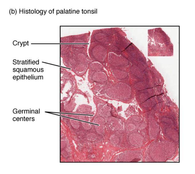
The mucosa-associated lymphoid tissue (MALT) nodule is a crucial component of the immune system, located within the mucosal linings of the gastrointestinal tract. This histological image, captured at a magnification of ×40, provides a detailed view of the MALT nodule’s structure, particularly within the small intestine, highlighting its role in local immune defense. Examining this micrograph offers valuable insights into how the body protects itself from pathogens encountered through the digestive system.

Peyer’s patches: Peyer’s patches are organized lymphoid follicles found in the mucosa of the small intestine, serving as a primary site for immune surveillance. They contain lymphocytes that detect and respond to antigens in the gut, initiating an immune response to protect against infections.
Mucosa: The mucosa is the innermost layer of the intestinal wall, consisting of epithelial cells and underlying lymphoid tissue, including Peyer’s patches. It acts as a barrier that absorbs nutrients while housing immune cells to monitor and combat pathogens entering through the gut.
Anatomical Structure of MALT Nodules
The MALT nodule, particularly Peyer’s patches, is an integral part of the gut-associated lymphoid tissue (GALT). These structures are strategically positioned to monitor the intestinal environment.
- Peyer’s patches form clusters of lymphoid follicles, rich in T and B lymphocytes, which are essential for antigen recognition.
- These patches are embedded within the mucosa, which provides a moist surface for nutrient absorption and immune activity.
- The lymphoid tissue within Peyer’s patches includes germinal centers where B cells mature, enhancing localized immunity.
- This arrangement allows the MALT to effectively sample antigens from the gut lumen, triggering an immune response when needed.
The MALT’s location in the intestine underscores its role in maintaining a balanced gut microbiome.
Microscopic Features and Functionality
This histological image, magnified at ×40, reveals the intricate cellular organization of the MALT nodule. The clarity of the micrograph aids in understanding its physiological roles.
- Peyer’s patches appear as dense, darkly stained areas, indicating high lymphocyte concentration, crucial for pathogen detection.
- The mucosa’s epithelial layer is lined with microvilli, increasing surface area for absorption while supporting immune cells beneath.
- Specialized M cells within the mucosa transport antigens to Peyer’s patches, facilitating immune activation.
- This microscopic structure ensures continuous monitoring of the intestinal contents, protecting against bacterial invasion.
The interplay of these components highlights the MALT’s efficiency in gut immunity.
Physical Characteristics and Clinical Relevance
The MALT nodule’s physical properties reflect its adaptive function within the intestinal wall. Its size and density can vary depending on immune activity levels.
- Peyer’s patches can enlarge during infections, reflecting increased lymphocyte recruitment to combat pathogens.
- The mucosa’s thin yet resilient layer adapts to mechanical stress from food passage while harboring immune defenses.
- Inflammation of Peyer’s patches may occur in conditions like Crohn’s disease, indicating an overactive immune response.
- Understanding these features aids in diagnosing gastrointestinal immune disorders, emphasizing the MALT’s clinical importance.
The MALT’s ability to regenerate lymphoid tissue supports its long-term protective role.
Conclusion
The histological image of the MALT nodule provides a detailed perspective on its vital role in gut immunity, particularly through Peyer’s patches and the mucosa. These structures work together to detect, process, and neutralize pathogens, safeguarding the digestive system. This exploration of the MALT nodule’s microstructure lays a strong foundation for studying its functions and addressing related health concerns effectively.

