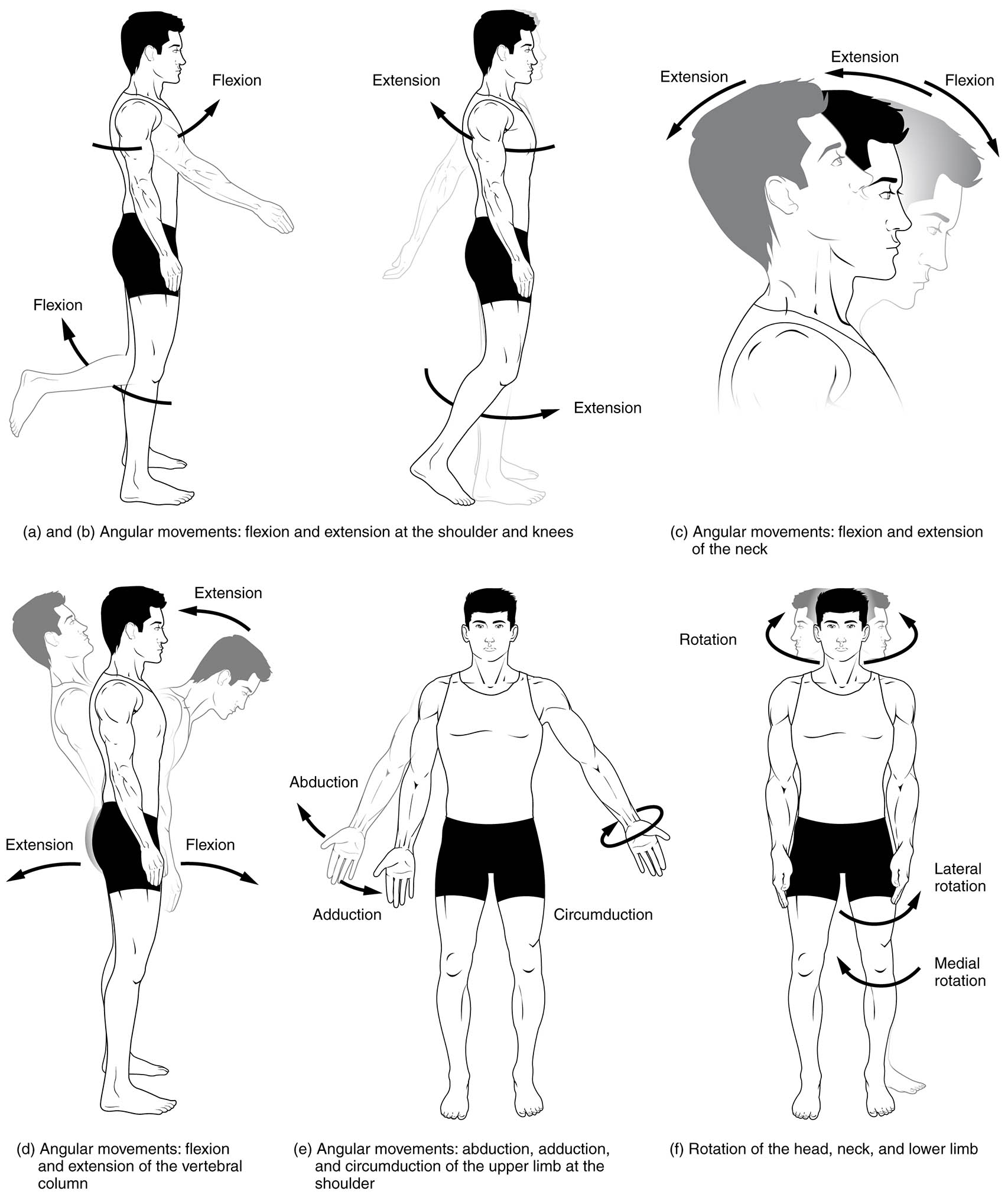
The human body’s ability to move is facilitated by the versatile synovial joints, which allow a wide range of motions essential for daily activities. This diagram illustrates key movements such as flexion, extension, abduction, adduction, circumduction, and rotation, categorized by their planes and joint involvement, providing a detailed view of anatomical mobility. Exploring this image offers a deeper understanding of how these movements contribute to the body’s functionality and coordination.
Labelled Parts Explanation
- Flexion The flexion movement involves bending a joint to decrease the angle between two bones, occurring in the sagittal plane, such as bending the elbow or knee. It is a fundamental motion for activities like walking or lifting, enabled by flexor muscles.
- Extension The extension movement straightens a joint to increase the angle between bones, also in the sagittal plane, such as straightening the arm or leg. It is critical for returning to an upright position or pushing movements, driven by extensor muscles.
- Abduction The abduction movement involves moving a limb or digits away from the body’s midline in the coronal plane, such as raising the arm sideways or spreading fingers. It is essential for reaching or balancing, facilitated by abductor muscles.
- Adduction The adduction movement brings a limb or digits toward or across the body’s midline in the coronal plane, such as lowering the arm or closing fingers. It supports actions like bringing objects closer or stabilizing the body, aided by adductor muscles.
- Circumduction The circumduction movement describes a circular motion of a limb or digits, combining flexion, adduction, extension, and abduction in sequence. It occurs at joints like the shoulder or hip, enabling complex motions like drawing a circle with the arm.
- Rotation The rotation movement involves turning a body part around its long axis, such as turning the head side to side or rotating the shoulder. It includes medial (internal) and lateral (external) rotation, supporting actions like looking over the shoulder or pivoting the leg.
Anatomical Overview of Body Movements
Synovial joints provide the structural foundation for the body’s diverse range of motions. This diagram categorizes movements by plane and joint, illustrating their anatomical basis.
- The flexion and extension occur in the sagittal plane, affecting joints like the elbow and knee.
- The abduction and adduction operate in the coronal plane, involving the shoulder and fingers.
- The circumduction combines multiple planes, seen at the hip and wrist.
- The rotation allows axial movements, notably at the neck and hip.
These movements reflect the joint’s adaptability to various activities.
Flexion and Extension in the Sagittal Plane
Flexion and extension are primary movements in the sagittal plane. Their mechanics support everyday tasks.
- The flexion bends joints like the hip or wrist, reducing the angle for actions like sitting.
- The extension straightens these joints, such as standing or extending the arm.
- These motions occur at synovial joints with hinge or ball-and-socket designs.
- Muscle groups like flexors and extensors coordinate these actions.
This plane is key to forward and backward motion.
Abduction and Adduction in the Coronal Plane
Abduction and adduction enable lateral movements in the coronal plane. Their function aids in spatial orientation.
- The abduction moves limbs outward, like raising the leg sideways.
- The adduction brings limbs inward, such as crossing the arms.
- These movements occur at joints with wide range, like the shoulder.
- Adductor and abductor muscles power these lateral shifts.
This plane supports side-to-side stability.
Circumduction as a Combined Motion
Circumduction involves a circular pattern, integrating multiple movements. Its complexity enhances joint versatility.
- The circumduction allows the arm to trace a circle, using sequential joint actions.
- It combines flexion, adduction, extension, and abduction in a smooth flow.
- This motion is prominent at ball-and-socket joints like the shoulder.
- It supports activities requiring circular reach, like throwing.
This movement showcases joint flexibility.
Rotation and Axial Movements
Rotation provides rotational freedom around an axis. Its variations support diverse head and limb actions.
- The rotation turns the head or limb, with medial rotation toward the midline.
- Lateral rotation moves the limb away, as in turning the foot outward.
- This occurs at pivot or ball-and-socket joints like the neck or hip.
- Rotator muscles drive these twisting motions.
This axis enhances directional control.
Physiological Importance of Joint Movements
These movements are essential for maintaining body function and mobility. Their coordination supports daily life.
- The flexion and extension facilitate walking and standing.
- The abduction and adduction aid in balance and reaching.
- The circumduction and rotation enable complex tasks like sports.
- Joint health ensures these motions remain effective.
This versatility underpins physical capability.
Clinical Relevance of Body Movements
Understanding joint movements assists in diagnosing musculoskeletal issues. These actions are key clinical indicators.
- Limited flexion or extension may suggest arthritis or joint stiffness.
- Abnormal abduction or adduction can indicate nerve damage or muscle weakness.
- Reduced circumduction might reflect shoulder impingement.
- Rotation deficits could signal spinal or hip pathology.
This knowledge guides rehabilitation and treatment.
Conclusion
The movements of the body medical description diagram provides a detailed exploration of how flexion, extension, abduction, adduction, circumduction, and rotation enable the body’s mobility through synovial joints. By examining these movements across the sagittal, coronal, and axial planes, one gains insight into the anatomical and functional coordination that supports daily activities. This understanding serves as a foundation for studying musculoskeletal health and addressing related concerns, encouraging further exploration of joint mechanics and their role in maintaining an active lifestyle.

