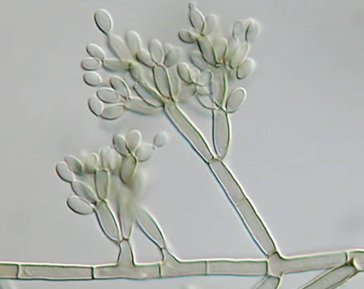
This high-resolution Differential Interference Contrast (DIC) micrograph captures the structural intricacies of Fonsecaea pedrosoi, a major fungal pathogen grown on modified Leonian’s agar. By visualizing the distinct arrangement of hyphae and conidia without the need for chemical staining, this image provides essential diagnostic clues for identifying the primary etiological agent of chromoblastomycosis, a debilitating chronic skin disease found in tropical regions.
Morphology and Identification of Fonsecaea pedrosoi
Fonsecaea pedrosoi is a dematiaceous fungus, meaning it naturally produces melanin, giving its colonies a dark olive-to-black appearance macroscopically. The image utilizes Differential Interference Contrast microscopy, a technique that enhances the contrast in unstained, transparent samples, giving the fungal structures a pseudo-3D, relief-like quality. This allows for a detailed examination of the fungal reproductive structures, which are critical for accurate laboratory identification. In the image, we observe septate hyphae—the branching filaments that make up the body of the fungus—giving rise to conidiophores (specialized stalks).
The reproductive morphology of Fonsecaea is known for being polymorphic, meaning it can display different arrangements of spores (conidia). The primary form visible here typically involves a sympodial arrangement, where conidia develop on the upper portion of the conidiophore. Secondary and tertiary conidia often form in short chains or clusters, creating a complex, tree-like structure. Microbiologists look for these specific branching patterns to distinguish F. pedrosoi from other closely related black molds found in environmental samples.
This fungus is saprophytic, thriving in soil, rotten wood, and decomposing plant matter. Its ability to survive in harsh environmental conditions is partly due to its melanized cell walls, which offer protection against ultraviolet radiation and enzymatic lysis. This resilience translates to its persistence in human host tissues upon infection. In the laboratory, modified Leonian’s agar is often used to stimulate this characteristic sporulation, as standard media may sometimes yield only sterile vegetative hyphae.
Key characteristics of Fonsecaea pedrosoi include:
- Pigmentation: Melanin production results in dark-colored hyphae and conidia.
- Growth Rate: It is a slow-growing fungus, often taking weeks to mature in culture.
- Habitat: Commonly found in tropical and subtropical soil and vegetation.
- Thermotolerance: Capable of growing at human body temperature, facilitating infection.
Clinical Significance: Chromoblastomycosis
Fonsecaea pedrosoi is the most common cause of chromoblastomycosis, a chronic, progressive fungal infection affecting the skin and subcutaneous tissues. The disease is primarily an occupational hazard for agricultural workers, lumberjacks, and individuals in rural tropical areas who are frequently exposed to soil and plant material. Infection occurs through traumatic inoculation—essentially, the fungus is implanted into the skin via a thorn prick, splinter, or minor cut. Because the fungus resides on plants, walking barefoot or handling wood without protection significantly increases the risk of exposure.
The clinical presentation begins slowly, often starting as a small, itchy papule or nodule at the site of trauma. Over months or years, the lesion expands and evolves. The hallmark presentation involves the development of verrucous (wart-like) or cauliflower-like masses that can become ulcerated and crusted. These lesions are typically painless but can be itchy, leading to scratching that spreads the infection to surrounding skin (autoinoculation). In severe, long-standing cases, the lymphatic system may become obstructed, leading to secondary lymphedema (swelling) that can resemble elephantiasis. Secondary bacterial infections are also common due to the compromised skin barrier.
A definitive diagnosis involves more than just culturing the organism; it requires the identification of pathognomonic structures in tissue samples. When skin scrapings or biopsies from a patient are examined under a microscope, the presence of sclerotic bodies (also known as Muriform cells or “copper pennies”) confirms the diagnosis. These are thick-walled, dark brown, multi-celled structures that represent the vegetative form of the fungus within the tissue. They divide by internal septation rather than budding, distinguishing this condition from other fungal infections like blastomycosis.
Conclusion
The microscopic identification of Fonsecaea pedrosoi, as seen in this DIC image, is a vital step in connecting environmental exposure to clinical pathology. While the fungus displays delicate branching conidia in the laboratory, its manifestation as resilient sclerotic bodies in human tissue leads to the challenging and chronic nature of chromoblastomycosis. Understanding the morphology and ecology of this organism aids healthcare providers in early diagnosis and the implementation of antifungal therapies to manage this disfiguring disease.

