The male urethra is a unique and functionally versatile tube, serving as a common pathway for both the urinary and reproductive systems. This article provides a comprehensive overview of the male urethra sectional anatomy and its surrounding structures, highlighting its different segments and connections to various accessory glands. Understanding this intricate anatomy is crucial for comprehending urinary and reproductive health, as well as various conditions affecting these systems.
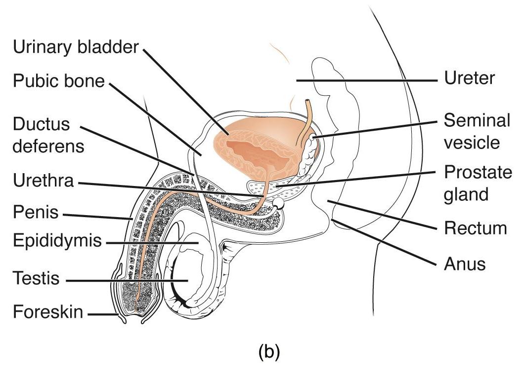
Urinary bladder: This is a muscular, hollow organ responsible for storing urine before it is expelled from the body. It sits in the pelvic cavity, posterior to the pubic bone.
Pubic bone: This is the most anterior bone of the pelvis, providing protection and attachment points for muscles and ligaments. The urinary bladder is located immediately posterior to it.
Ductus deferens: Also known as the vas deferens, these muscular tubes transport sperm from the epididymis to the ejaculatory duct. They ascend from the scrotum, loop over the ureters, and join the seminal vesicles.
Urethra: In males, this tube extends from the urinary bladder through the penis, serving as the exit pathway for both urine and semen. It has three main sections: prostatic, membranous, and spongy (penile) urethra.
Penis: This is the male external reproductive organ, containing the urethra and involved in sexual intercourse and urination. It is composed of erectile tissues that fill with blood during arousal.
Epididymis: This coiled tube sits on the posterior side of each testis, serving as a site for sperm maturation and storage. Sperm gain motility and fertilizing ability as they pass through here.
Testis: These are the primary male reproductive organs, located within the scrotum. They produce sperm (spermatogenesis) and male hormones, primarily testosterone.
Foreskin: Also known as the prepuce, this is a retractable fold of skin that covers the glans (head) of the penis. It may be surgically removed during circumcision.
Ureter: These tubes transport urine from the kidneys to the urinary bladder. They enter the bladder at its posterior-inferior aspect.
Seminal vesicle: These are a pair of glands located posterior to the urinary bladder, which secrete a viscous, alkaline fluid that forms a significant portion of semen. This fluid contains fructose for sperm nourishment and other components that enhance sperm motility.
Prostate gland: This gland is located inferior to the urinary bladder and surrounds the initial part of the urethra. It secretes a milky, slightly acidic fluid into the urethra, contributing to semen volume and sperm activation.
Rectum: This is the final section of the large intestine, ending at the anus. It is located posterior to the urinary bladder and prostate gland.
Anus: This is the external opening of the rectum, through which feces are expelled from the body. Its function is controlled by internal and external anal sphincters.
The male urethra represents a remarkable anatomical convergence, uniquely designed to serve both the urinary and reproductive systems. Unlike the female urethra, which is solely dedicated to urine transport, the male urethra provides a conduit for both urine and semen, necessitating intricate regulatory mechanisms to ensure proper function. Its extended length and passage through various pelvic structures and the penis make it susceptible to different conditions compared to its female counterpart. This sectional view offers a comprehensive look at the urethra’s pathway and its intimate relationships with key accessory reproductive glands and the urinary bladder.
The male urethra is typically divided into three distinct segments:
- Prostatic Urethra: This is the widest and most dilatable part, approximately 3-4 cm long, passing through the prostate gland. It receives ejaculatory ducts from the seminal vesicles and ductus deferens, as well as ducts from the prostate gland.
- Membranous Urethra: The shortest and narrowest part, about 1-2 cm long, this segment passes through the deep perineal pouch. It is surrounded by the external urethral sphincter, which is composed of voluntary skeletal muscle and provides conscious control over urination.
- Spongy (Penile) Urethra: The longest segment, roughly 15 cm, it passes through the corpus spongiosum of the penis and opens at the external urethral orifice. Ducts from the bulbourethral glands open into this part.
The prostate gland, strategically encircling the prostatic urethra, is particularly significant as its enlargement can directly impact urinary flow. Benign prostatic hyperplasia (BPH), a common condition in older men, involves the non-cancerous growth of the prostate gland. As the prostate enlarges, it compresses the urethra, leading to symptoms such as urinary frequency, urgency, weak stream, and incomplete bladder emptying. Severe cases can cause urinary retention and even kidney damage. Furthermore, the close proximity of the seminal vesicles and ductus deferens to the urethra underscores the urethra’s dual role. During ejaculation, sperm from the testes and epididymis travel through the ductus deferens, mix with fluids from the seminal vesicles and prostate gland to form semen, which then passes through the urethra. This intricate anatomical arrangement allows for coordinated function but also makes the male urinary and reproductive systems susceptible to interconnected health issues.

