The venous system of the lower limbs is a critical pathway for returning deoxygenated blood from the legs and feet to the heart, relying on a complex network of deep and superficial veins. This posterior view showcases the anatomical layout of these veins, highlighting their role in maintaining circulation against gravity with the aid of muscular pumps and one-way valves. Gaining insight into this structure enhances understanding of how the body sustains mobility and prevents circulatory stagnation.
Detailed Anatomy of Labeled Veins
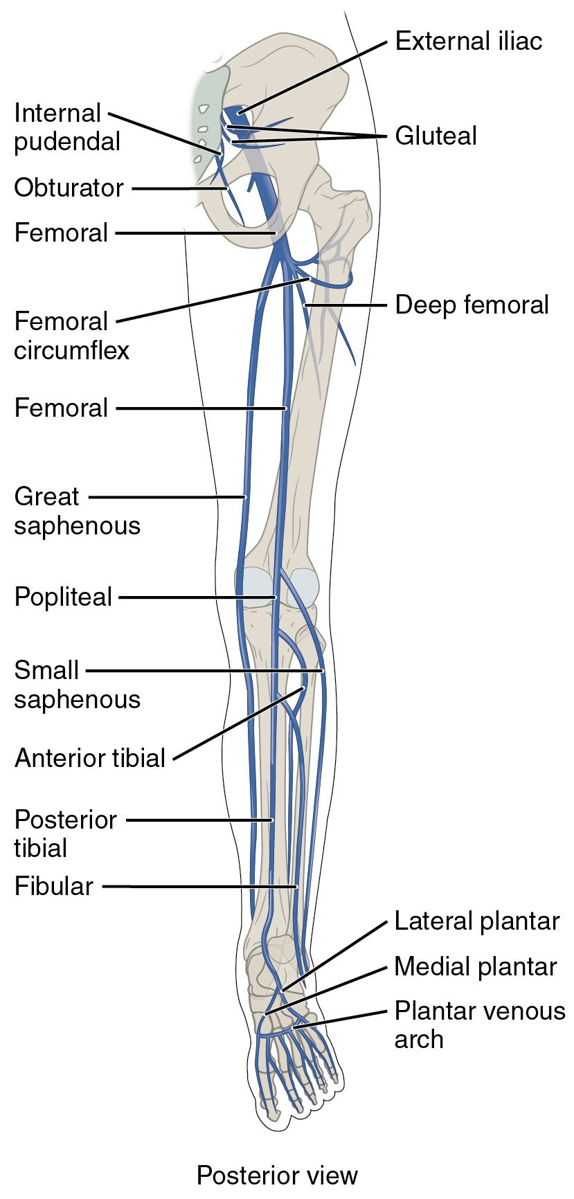
The following sections explore each labeled vein from the posterior perspective, detailing their courses and physiological contributions.
External iliac: The external iliac vein begins at the inguinal ligament as a continuation of the femoral vein, collecting blood from the lower limb. It merges with the internal iliac vein to form the common iliac, facilitating the transition of blood into the pelvic region.
Gluteal: The gluteal veins drain the gluteal muscles and surrounding tissues, running alongside the gluteal arteries to join the internal iliac vein. These veins are vital for circulation in the buttocks, supporting posture and physical movement.
Internal pudendal: The internal pudendal vein collects blood from the perineal region, including the external genitalia and anal area, following the pudendal artery. It contributes to the internal iliac vein, ensuring efficient venous return from the pelvic floor.
Obturator: The obturator vein drains the adductor muscles and hip joint, passing through the obturator canal to connect with the internal iliac vein. This vessel supports circulation in the medial thigh, aiding hip joint stability.
Femoral: The femoral vein is a major deep vein that starts as the popliteal vein and runs through the thigh, becoming the external iliac at the inguinal ligament. It receives blood from both deep and superficial tributaries, playing a central role in lower limb venous return.
Femoral circumflex: The femoral circumflex veins, including medial and lateral branches, drain the upper thigh and hip region, accompanying the circumflex arteries. They typically empty into the femoral or deep femoral veins, supporting circulation around the hip joint.
Deep femoral: The deep femoral vein, or profunda femoris, runs parallel to the deep femoral artery, draining the deeper thigh muscles. It joins the femoral vein, providing a key pathway for blood from the quadriceps and hamstrings.
Great saphenous: The great saphenous vein, the longest superficial vein, ascends medially from the dorsal venous arch to join the femoral vein at the saphenous opening. It drains the medial and anterior leg and thigh, often harvested for vascular grafts.
Popliteal: The popliteal vein forms behind the knee from the union of the anterior and posterior tibial veins, continuing as the femoral vein. It drains the knee joint and calf muscles, relying on valves to prevent backflow during movement.
Small saphenous: The small saphenous vein runs along the posterior leg from the lateral foot to the popliteal fossa, joining the popliteal vein. It drains the lateral and posterior leg, complementing the great saphenous in superficial circulation.
Anterior tibial: The anterior tibial vein drains the anterior compartment of the leg, including dorsiflexor muscles, and ascends to form the popliteal with the posterior tibial. Paired with its artery, it ensures blood return from the shin critical for foot movement.
Posterior tibial: The posterior tibial vein runs deep in the calf, draining the plantar foot and posterior leg muscles, joining the anterior tibial at the popliteal. It supports upward blood flow through calf muscle action and perforating veins.
Fibular: The fibular vein, or peroneal, accompanies the fibular artery in the lateral calf, draining deep tissues and merging with the posterior tibial. This vein aids circulation in the lateral leg, supporting balance and peroneal muscle function.
Lateral plantar: The lateral plantar vein drains the lateral sole of the foot, accompanying the lateral plantar artery and joining the plantar venous arch. It supports weight-bearing areas, contributing to overall foot venous drainage.
Medial plantar: The medial plantar vein collects blood from the medial sole, running with the medial plantar artery to enter the plantar venous arch. This vein is crucial for maintaining circulation in the arch of the foot during locomotion.
Plantar venous arch: The plantar venous arch forms a network under the foot, receiving digital and plantar veins before continuing as the posterior tibial. It acts as a reservoir for blood during standing, with muscle pumps aiding return flow.
Physiological Role of Lower Limb Veins
The posterior view reveals veins designed to overcome gravitational challenges, utilizing muscular contractions and valves for effective circulation.
- Deep veins like the femoral and popliteal carry the majority of venous blood, benefiting from the skeletal muscle pump during activities such as walking.
- Superficial veins, including the great and small saphenous, provide an alternative route, connecting to deep veins via perforators for heat regulation.
- Valves ensure unidirectional flow, preventing reflux and maintaining pressure gradients toward the heart.
- Blood from the foot ascends through the plantar arch and tibial veins, merging at key junctions to enter the thigh and pelvis efficiently.
Clinical Relevance and Preventive Measures
Understanding venous anatomy from the posterior perspective aids in addressing common circulatory issues and promoting health.
- Varicose veins often affect the great saphenous, leading to bulging and discomfort due to valve failure and increased pressure.
- Deep vein thrombosis may involve the femoral or popliteal veins, where clots from immobility pose a risk of pulmonary embolism.
- Treatments such as compression therapy or surgical vein stripping target problematic veins while preserving deep circulation.
- Regular exercise and leg elevation enhance venous return, mimicking the natural pump to reduce swelling and improve flow.
Integration with Systemic Circulation
The posterior veins connect with the broader circulatory system, supporting overall physiological balance.
- Blood from the external iliac joins the inferior vena cava via the common iliac, delivering it to the right atrium for oxygenation.
- Hormones like adrenaline from the adrenal glands influence vascular tone, affecting fluid dynamics in these veins.
- Prolonged standing or pregnancy can increase pressure on iliac veins, causing temporary edema that movement can alleviate.
- Lymphatic vessels alongside these veins manage interstitial fluid, complementing venous drainage and immune responses.
In conclusion, the major veins of the lower limbs, as seen in the posterior view, form a robust network that adapts to the demands of movement and posture. A thorough grasp of their anatomy encourages practices that sustain healthy circulation, fostering long-term vascular well-being.

