The venous system of the lower limb is a sophisticated network designed to return deoxygenated blood to the heart, efficiently managing flow against gravity. This flow chart illustrates the hierarchical structure of major veins, highlighting their roles in collecting and transporting blood from the foot to the central circulation. Exploring this diagram provides a clear understanding of how these vessels collaborate to maintain circulatory health and support physical activity.
Detailed Anatomy of Labeled Veins
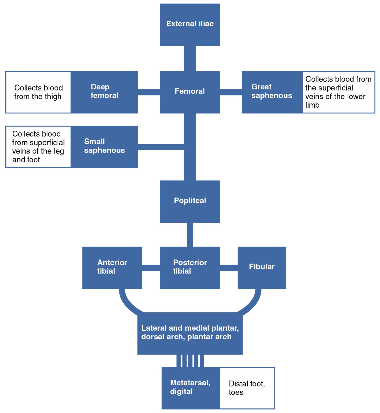
The following sections outline each labeled vein, explaining its function and position within the venous flow.
External iliac: The external iliac vein serves as the upper boundary of the lower limb venous system, receiving blood from the femoral vein at the inguinal ligament. It merges with the internal iliac vein to form the common iliac, directing blood toward the inferior vena cava.
Deep femoral: The deep femoral vein, or profunda femoris, collects blood from the deeper thigh muscles and joins the femoral vein. It plays a crucial role in draining the posterior and medial thigh regions during movement.
Femoral: The femoral vein is a major deep vein that runs through the thigh, receiving blood from the deep femoral and great saphenous veins. It transitions into the external iliac vein at the inguinal ligament, serving as a central conduit for lower limb drainage.
Great saphenous: The great saphenous vein is the longest superficial vein, collecting blood from the superficial veins of the lower limb and joining the femoral vein. It originates from the dorsal venous arch and drains the medial leg and thigh.
Small saphenous: The small saphenous vein gathers blood from the superficial veins of the leg and foot, running along the posterior calf to join the popliteal vein. It provides an essential superficial drainage route for the lateral and posterior leg.
Popliteal: The popliteal vein forms behind the knee from the union of the anterior and posterior tibial veins, continuing as the femoral vein. It collects blood from the calf and knee region, relying on valves to prevent backflow.
Anterior tibial: The anterior tibial vein drains the anterior compartment of the leg, including the dorsiflexor muscles, and merges with the posterior tibial to form the popliteal. It ensures blood return from the shin, supporting foot movement.
Posterior tibial: The posterior tibial vein runs deep in the calf, collecting blood from the plantar foot and posterior leg muscles, and joins the anterior tibial to form the popliteal. It facilitates upward flow through calf muscle contractions.
Fibular: The fibular vein, or peroneal, drains the lateral compartment of the calf and merges with the posterior tibial vein. It supports circulation in the peroneal muscles, aiding balance and stability.
Lateral and medial plantar, dorsal arch, plantar arch: The lateral and medial plantar, dorsal arch, plantar arch veins form a network under the foot, collecting blood from the metatarsal and digital regions. These veins converge to feed into the posterior tibial, acting as a reservoir during weight-bearing.
Metatarsal, distal foot, toes: The metatarsal, distal foot, toes veins drain the toes and metatarsal area, feeding into the plantar and dorsal arches. They serve as the initial collection points for deoxygenated blood in the distal lower limb.
Physiological Role of Lower Limb Veins
This flow chart reveals how veins work together to ensure efficient blood return, supported by muscular and valvular mechanisms.
- The external iliac and femoral veins form the primary deep pathway, handling the bulk of venous return from the thigh and leg.
- Superficial veins like the great saphenous and small saphenous collect blood from the skin and subcutaneous tissues, connecting to deep veins via perforators.
- The popliteal and tibial veins rely on the calf muscle pump to propel blood upward, with valves preventing reflux.
- Blood from the metatarsal, distal foot, toes ascends through interconnected arches, ensuring comprehensive drainage from the foot.
Clinical Relevance and Health Practices
Understanding this venous flow aids in recognizing potential circulatory issues and promoting wellness.
- Varicose veins often involve the great saphenous, resulting from valve incompetence and increased pressure, leading to visible bulging.
- Deep vein thrombosis can affect the femoral or popliteal veins, where clots from prolonged immobility risk pulmonary embolism.
- Compression stockings enhance flow in the tibial and saphenous veins, mimicking the muscle pump to reduce swelling.
- Regular walking stimulates the popliteal and fibular veins, improving overall venous return and preventing stagnation.
Integration with Systemic Circulation
The lower limb venous system connects seamlessly with the body’s broader circulatory network, supporting homeostasis.
- Blood from the external iliac enters the inferior vena cava, delivering it to the right atrium for reoxygenation.
- Hormones like cortisol from the adrenal glands influence vascular tone, affecting fluid dynamics in the femoral and deep femoral veins.
- Pregnancy or prolonged standing increases pressure on the iliac veins, potentially causing edema that movement can mitigate.
- Lymphatic vessels parallel these veins, managing interstitial fluid and supporting immune function alongside venous return.
In conclusion, the major veins of the lower limb, as depicted in this flow chart, form an intricate system that ensures efficient blood return to the heart. Appreciating their structure and function encourages habits that sustain healthy circulation, contributing to long-term vascular health.

