The human body’s venous system plays a crucial role in returning deoxygenated blood from the peripheries back to the heart, ensuring efficient circulation and nutrient distribution. This intricate network of veins, illustrated in the anterior view of major systemic veins, highlights key pathways that support vital physiological functions, from oxygen transport to waste removal.
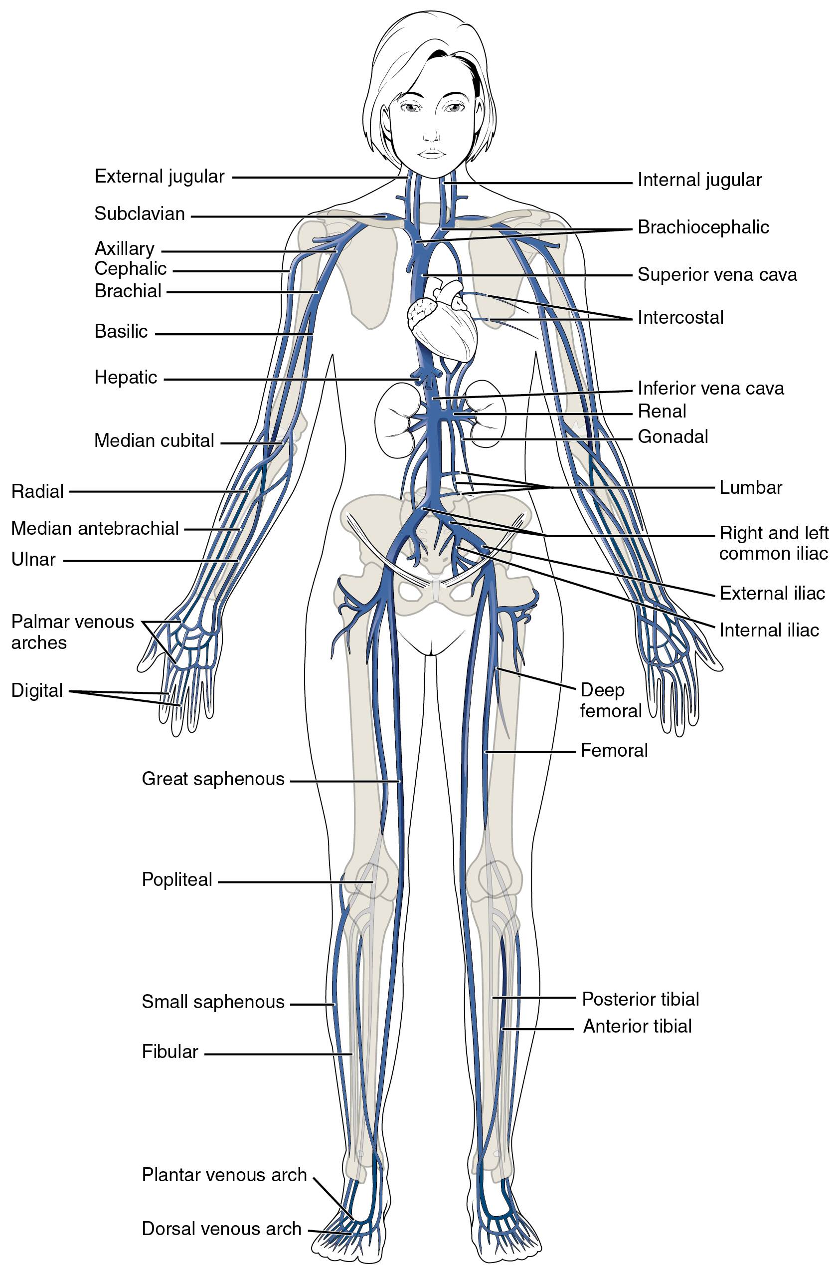
External jugular The external jugular vein drains blood from the superficial regions of the head and neck, including the scalp and face. It descends along the side of the neck and joins the subclavian vein to form the brachiocephalic vein.
Internal jugular The internal jugular vein collects blood from the brain, deep face, and neck structures, running parallel to the common carotid artery. It merges with the subclavian vein to create the brachiocephalic vein, playing a key role in cerebral venous drainage.
Subclavian The subclavian vein receives blood from the upper limb and parts of the thorax, located beneath the clavicle. It unites with the internal jugular vein to form the brachiocephalic vein, facilitating blood return from the arms and shoulders.
Brachiocephalic The brachiocephalic vein is formed by the union of the subclavian and internal jugular veins, one on each side of the body. These veins converge to create the superior vena cava, channeling blood from the head, neck, and upper limbs to the heart.
Axillary The axillary vein drains the upper arm, shoulder, and pectoral regions, continuing from the brachial vein. It transitions into the subclavian vein at the lateral border of the first rib, essential for venous return from the armpit area.
Cephalic The cephalic vein is a superficial vein of the upper limb, running along the lateral aspect from the hand to the shoulder. It empties into the axillary vein, commonly used for venipuncture due to its accessibility.
Brachial The brachial vein accompanies the brachial artery in the upper arm, draining deep tissues. It joins with the basilic vein to form the axillary vein, supporting blood flow from the elbow region upward.
Basilic The basilic vein is a superficial vein on the medial side of the arm, starting from the dorsal venous network of the hand. It ascends to join the brachial vein, forming the axillary vein, and is often utilized in medical procedures like IV insertions.
Hepatic The hepatic veins drain deoxygenated blood from the liver into the inferior vena cava. They play a vital role in processing blood from the digestive tract, ensuring metabolites are transported efficiently.
Median cubital The median cubital vein connects the cephalic and basilic veins in the antecubital fossa. It is a common site for blood draws and IV access due to its prominence and stability.
Radial The radial vein runs along the lateral forearm, draining the hand and wrist areas. It unites with the ulnar vein to form the brachial vein, aiding in venous return from the thumb side.
Median antebrachial The median antebrachial vein drains the anterior forearm, located centrally. It typically joins the basilic or median cubital vein, contributing to superficial drainage of the arm.
Ulnar The ulnar vein accompanies the ulnar artery on the medial forearm, draining the little finger side. It merges with the radial vein to create the brachial vein, essential for hand and forearm circulation.
Palmar venous arches The palmar venous arches are networks in the palm, including superficial and deep arches that drain the fingers. They connect to forearm veins, facilitating blood return from hand activities.
Digital Digital veins drain the fingers and toes, collecting blood from phalanges. They feed into venous arches, ensuring efficient drainage from extremities.
Superior vena cava The superior vena cava receives blood from the upper body, formed by brachiocephalic veins. It empties into the right atrium, crucial for systemic circulation.
Intercostal Intercostal veins drain the intercostal spaces between ribs, collecting blood from chest walls. They empty into the azygos system or directly into the superior vena cava.
Inferior vena cava The inferior vena cava is the largest vein, carrying blood from the lower body to the heart. It ascends through the abdomen, receiving tributaries from viscera and limbs.
Renal Renal veins drain the kidneys, with the right shorter than the left due to anatomical positioning. They enter the inferior vena cava, vital for filtering blood wastes.
Gonadal Gonadal veins drain the ovaries or testes, with the right entering the inferior vena cava and left into the renal vein. They transport blood from reproductive organs, influencing hormonal circulation.
Lumbar Lumbar veins drain the posterior abdominal wall and vertebral column. They empty into the inferior vena cava, supporting spinal and muscular drainage.
Right and left common iliac The common iliac veins form from external and internal iliac veins, one on each side. They unite to create the inferior vena cava, channeling lower limb and pelvic blood.
External iliac The external iliac vein drains the lower limb, continuing from the femoral vein. It joins the internal iliac to form the common iliac, key for leg circulation.
Internal iliac The internal iliac vein collects blood from pelvic organs like bladder and rectum. It merges with the external iliac, ensuring pelvic venous return.
Deep femoral The deep femoral vein drains deep thigh muscles, running alongside the profunda femoris artery. It joins the femoral vein, enhancing lower limb drainage.
Femoral The femoral vein is the main vein of the thigh, continuing from the popliteal. It becomes the external iliac at the inguinal ligament, critical for leg blood return.
Great saphenous The great saphenous vein is the longest superficial vein, from foot to groin. It drains into the femoral vein, often involved in varicose conditions.
Popliteal The popliteal vein is behind the knee, formed by tibial veins. It continues as the femoral vein, facilitating calf to thigh transition.
Small saphenous The small saphenous vein is superficial on the posterior leg, from foot to knee. It empties into the popliteal vein, aiding lateral ankle drainage.
Fibular The fibular vein, also known as peroneal, drains lateral leg compartments. It joins posterior tibial veins, supporting lower leg circulation.
Posterior tibial The posterior tibial vein drains the posterior leg and foot plantar surface. It unites with anterior tibial to form popliteal, essential for calf muscles.
Anterior tibial The anterior tibial vein drains the anterior leg compartment. It merges with posterior tibial, contributing to overall leg venous flow.
Plantar venous arch The plantar venous arch drains the sole of the foot. It connects to tibial veins, aiding in weight-bearing drainage.
Dorsal venous arch The dorsal venous arch drains the top of the foot. It feeds into saphenous veins, crucial for foot circulation.
Understanding the Systemic Venous System
The systemic veins form an extensive network responsible for returning deoxygenated blood to the heart after it has delivered oxygen and nutrients to tissues. This system contrasts with the pulmonary veins, which carry oxygenated blood.
- Anatomical Overview: The veins are categorized into superficial and deep types, with superficial ones visible under the skin and deep ones accompanying arteries. This dual structure ensures redundancy and efficiency in blood return, preventing pooling and maintaining pressure gradients.
- Upper Body Drainage: Veins like the jugulars and subclavians handle head and arm blood, converging into the superior vena cava. This pathway is vital for rapid drainage from brain tissues to avoid increased intracranial pressure.
- Lower Body and Abdominal Drainage: The inferior vena cava collects from legs, pelvis, and organs via iliacs and viscerals. It supports filtration through kidneys and liver, integrating with digestive processes.
Physiological Functions of Major Veins
Veins contain valves to prevent backflow, relying on muscle contractions and respiration for propulsion. This one-way flow is essential for circulatory homeostasis.
- Role in Circulation: Systemic veins transport hormones, such as those from the thyroid gland including T3 and T4, throughout the body. They also carry carbon dioxide and wastes to lungs and kidneys for elimination.
- Clinical Relevance: Understanding vein locations aids in procedures like central line placements in the subclavian or femoral veins. Variations, such as accessory veins, can impact surgical outcomes.
- Pathway Integration: The hepatic veins, for instance, link portal and systemic circulations, metabolizing nutrients before heart return.
Comparative Anatomy and Variations
Human venous anatomy shares similarities with other mammals but features unique adaptations for bipedalism, like robust leg valves. Variations occur, such as duplicated inferior vena cava in some individuals.
- Embryological Development: Veins develop from cardinal veins in embryos, with anomalies leading to conditions like persistent left superior vena cava. This knowledge informs congenital defect diagnoses.
- Gender and Age Differences: Vein diameters may vary by gender, with hormonal influences affecting elasticity in females. Aging reduces valve competence, increasing risks like deep vein thrombosis.
In summary, the major systemic veins represent a sophisticated system integral to human physiology, adapting to demands of upright posture and activity. Mastery of this anatomy enhances appreciation of circulatory dynamics and informs medical interventions for optimal health.

