The aorta, the body’s largest artery, serves as the central highway for distributing oxygenated blood to every region, with its major branches playing a pivotal role. This flow chart outlines the distribution of these branches into the thoracic and abdominal regions, illustrating how they supply vital organs and tissues with essential nutrients and oxygen.
Aorta The aorta originates from the left ventricle, carrying oxygenated blood throughout the body. It divides into distinct regions, each with specialized branches for systemic circulation.
Ascending aorta This initial segment rises from the heart, distributing blood to the coronary arteries. It supports the heart muscle with a steady oxygen supply.
Aortic arch Curving between the ascending and descending aorta, it supplies the head, neck, and upper limbs. It gives rise to the brachiocephalic, left common carotid, and left subclavian arteries.
Descending aorta This major portion extends downward, delivering blood to the chest and abdomen. It is subdivided into thoracic and abdominal segments for targeted distribution.
Thoracic aorta Located in the chest, it supplies blood to the lungs, esophagus, and chest wall. Its branches support respiratory and structural functions.
Abdominal aorta Running through the abdomen, it provides blood to digestive organs, kidneys, and lower limbs. It branches into arteries like the celiac trunk and renal arteries.
Coronary arteries Branching from the ascending aorta, they supply the heart muscle. They ensure the myocardium receives oxygen for continuous pumping.
Brachiocephalic artery Arising from the aortic arch, it supplies the right arm and right side of the head and neck. It splits into the right subclavian and right common carotid arteries.
Left common carotid artery Originating from the aortic arch, it delivers blood to the left side of the head and neck. It supports brain and facial tissue oxygenation.
Left subclavian artery This aortic arch branch supplies the left arm and shoulder. It ensures blood flow for upper limb movement and function.
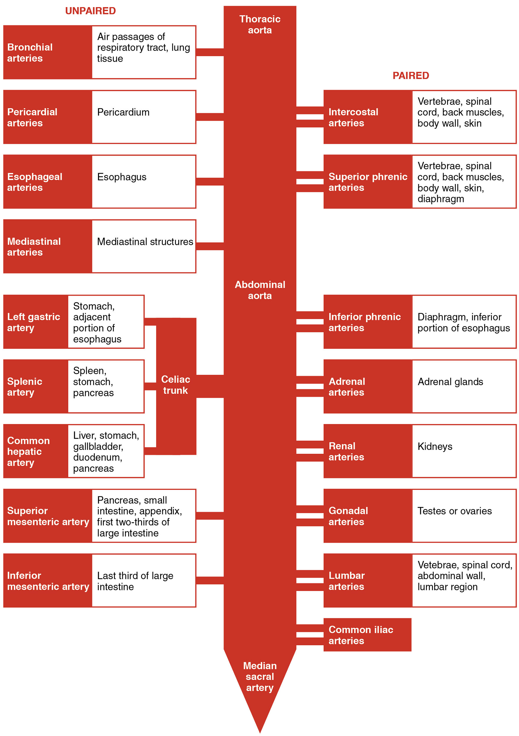
Pericardial arteries From the thoracic aorta, they supply the pericardium around the heart. They maintain the heart’s protective sac and structural health.
Bronchial arteries These thoracic aorta branches nourish the lungs. They support pulmonary tissue and bronchial function.
Esophageal arteries Originating from the thoracic aorta, they supply the esophagus. They ensure the esophagus remains viable for food transport.
Posterior intercostal arteries These thoracic aorta branches feed the intercostal muscles and chest wall. They aid in respiration by supporting rib movement.
Subcostal arteries Also from the thoracic aorta, they supply the lower chest wall and upper abdomen. They support the diaphragm and adjacent tissues.
Celiac trunk Branching from the abdominal aorta, it supplies the stomach, liver, and spleen. It facilitates digestion and blood filtration.
Superior mesenteric artery This abdominal aorta branch feeds the small intestine and part of the large intestine. It ensures nutrient absorption and gastrointestinal health.
Renal arteries Arising from the abdominal aorta, they supply the kidneys. They deliver blood for filtration and blood pressure regulation.
Inferior mesenteric artery This abdominal aorta branch serves the lower large intestine. It supports waste elimination and lower digestive function.
Common iliac arteries Splitting from the abdominal aorta, they supply the pelvis and lower limbs. They ensure blood flow to the legs and pelvic organs.
Structure of the Aorta and Its Initial Branches
The aorta begins its journey with the ascending aorta, setting the stage for blood distribution. Its early branches are tailored for critical functions.
- The ascending aorta gives rise to the coronary arteries to nourish the heart.
- The aortic arch curves to supply the upper body with key arteries.
- Elastic fibers in these segments absorb cardiac pressure waves.
- The design ensures high-pressure flow to meet systemic needs.
- Variations in branching can impact surgical approaches.
Thoracic Region Arterial Distribution
The thoracic aorta and its branches support the chest’s vital organs and structures. Its network adapts to respiratory and circulatory demands.
- Pericardial arteries protect the heart’s outer layer.
- Bronchial arteries sustain lung tissue health.
- Esophageal arteries ensure esophageal integrity.
- Posterior intercostal arteries aid chest wall movement.
- Subcostal arteries bridge the chest and abdomen.
Abdominal Region Arterial Supply
The abdominal aorta extends the aorta’s reach, feeding major abdominal organs. Its branches are crucial for digestion and filtration.
- The celiac trunk supports upper digestive functions.
- Superior mesenteric artery nourishes the intestines.
- Renal arteries ensure kidney efficiency.
- Inferior mesenteric artery serves the lower colon.
- Common iliac arteries reach the pelvis and legs.
Physiological Role of Aortic Branches
The major branches of the aorta maintain blood pressure and organ perfusion. Their function supports overall homeostasis.
- Coronary arteries prevent heart muscle ischemia.
- Arch branches like the brachiocephalic artery sustain brain and arm flow.
- Thoracic branches adjust to respiratory changes.
- Abdominal branches respond to digestive and excretory needs.
- This system’s adaptability prevents tissue hypoxia.
Clinical Significance of Aortic Branches
Understanding these branches aids in diagnosing and treating vascular conditions. Their anatomy guides medical interventions.
- Aneurysms in the aortic arch can affect upper body circulation.
- Renal arteries blockages may lead to hypertension.
- Thoracic aorta issues can compress lung or esophageal structures.
- Abdominal aortic aneurysms pose a rupture risk.
- Imaging techniques map these arteries for surgical planning.
The aorta and its major branches form an intricate network, delivering oxygenated blood to the thoracic and abdominal regions. This efficient distribution system ensures organs like the heart, lungs, and kidneys function optimally, providing a foundation for exploring cardiovascular health and addressing related challenges.

