The lower limb’s arterial network is essential for delivering oxygen-rich blood to support movement and tissue health, with the posterior view revealing the critical pathways. This image highlights the major arteries from the thigh to the foot, offering a comprehensive look at how circulation sustains the back of the leg, making it a valuable resource for anatomical study.
Exploring the Key Arteries in the Posterior View
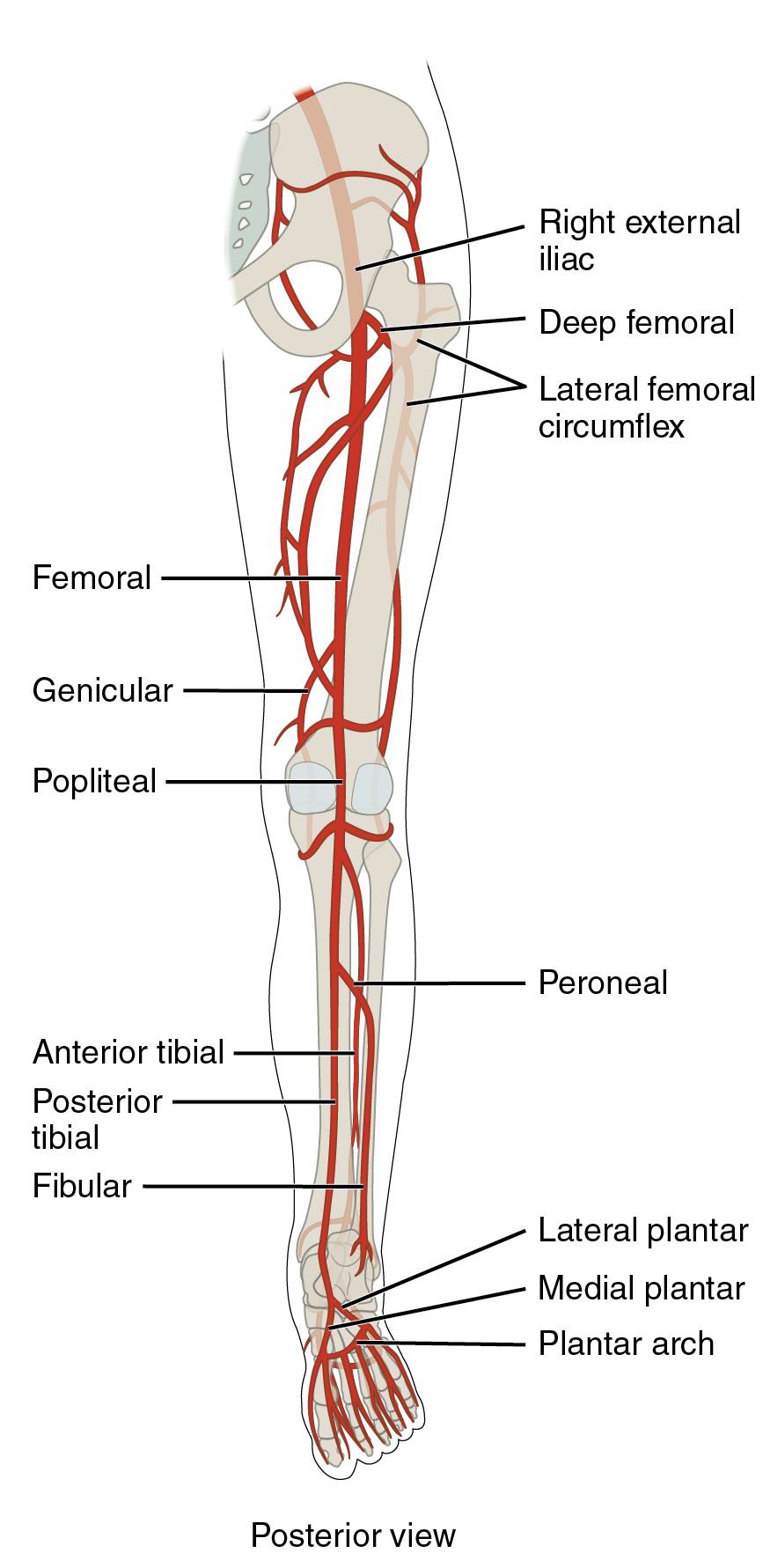
The labeled arteries in this image map the blood supply to the lower limb from a posterior perspective. Each vessel plays a distinct role in ensuring adequate perfusion to the muscles, joints, and tissues.
Right external iliac The right external iliac artery branches from the common iliac, transitioning into the femoral artery below the inguinal ligament. It serves as the primary route for blood flow to the right lower limb, supporting pelvic and thigh circulation.
Deep femoral The deep femoral artery branches from the femoral, supplying the deep muscles of the thigh and hip joint. It provides a robust collateral network, enhancing blood flow to the posterior thigh region.
Lateral femoral circumflex The lateral femoral circumflex artery arises from the deep femoral, encircling the femur to supply the lateral thigh muscles. It supports hip abduction and contributes to thigh stability during movement.
Femoral The femoral artery, a continuation of the external iliac, runs down the anterior thigh but is visible posteriorly as it transitions. It supplies the thigh muscles and serves as a key pulse point in the groin.
Genicular The genicular arteries branch from the femoral and popliteal, forming an anastomotic network around the knee. They ensure blood supply to the knee joint, supporting mobility and joint health.
Popliteal The popliteal artery continues from the femoral behind the knee, supplying the knee joint and posterior calf muscles. It bifurcates into the anterior and posterior tibial arteries, critical for lower leg circulation.
Peroneal The peroneal artery, also known as the fibular artery, branches from the posterior tibial, supplying the lateral leg and ankle. It provides collateral circulation, supporting the peroneal muscles and ankle stability.
Anterior tibial The anterior tibial artery branches from the popliteal, running along the anterior leg but visible posteriorly as it originates. It supplies the shin muscles and continues as the dorsalis pedis artery.
Posterior tibial The posterior tibial artery, a major popliteal branch, runs along the back of the leg, supplying the calf muscles and sole of the foot. It contributes to the plantar arch, ensuring foot perfusion.
Fibular The fibular artery, synonymous with the peroneal, branches from the posterior tibial, supplying the lateral leg. It enhances blood flow to the fibular region, supporting lateral ankle stability.
Lateral plantar The lateral plantar artery branches from the posterior tibial, supplying the lateral sole of the foot. It contributes to the plantar arch, supporting the foot’s weight-bearing surface.
Medial plantar The medial plantar artery, also from the posterior tibial, supplies the medial sole of the foot. It nourishes the plantar muscles and contributes to arch stability.
Plantar arch The plantar arch is formed by the lateral and medial plantar arteries, supplying the toes and sole. It ensures robust circulation to the weight-bearing plantar surface.
The Pathway of Lower Limb Arteries
Blood flow to the lower limb begins with the abdominal aorta, following a structured route visible in the posterior view. This pathway adapts to the leg’s posterior anatomical demands.
- The aorta bifurcates into the common iliac arteries, with the right external iliac continuing the flow.
- The femoral artery transitions into the popliteal behind the knee, adapting to the posterior compartment.
- The popliteal artery splits into the posterior tibial and peroneal arteries for distal circulation.
- The plantar arch completes the network, ensuring toe and sole perfusion.
Clinical Relevance of Posterior Arteries
Understanding these arteries aids in diagnosing vascular conditions from a posterior perspective. Their positions guide clinical interventions and assessments.
- The popliteal artery is a common site for aneurysm detection due to its posterior location.
- The posterior tibial pulse assesses foot perfusion, vital in peripheral artery disease.
- Genicular arteries support knee surgeries by providing collateral flow.
- Doppler ultrasound evaluates flow in the posterior tibial and peroneal arteries.
Physiological Roles in Lower Limb Function
These arteries adapt to physical demands, ensuring oxygen delivery to the posterior leg. Their responsiveness enhances mobility and endurance.
- The deep femoral and popliteal arteries dilate during exercise, boosting posterior thigh and calf performance.
- The plantar arch regulates blood flow to support standing and walking on the sole.
- Hormonal changes, such as adrenaline release, influence vascular tone in the posterior limb.
- Anastomoses around the knee protect against ischemic events in the posterior compartment.
In conclusion, the posterior view of the lower limb’s major arteries reveals a resilient network supporting movement and stability. Mastering this anatomy enhances clinical practice and deepens appreciation for circulatory efficiency.

