The lower limbs rely on a sophisticated arterial network to deliver oxygen-rich blood from the heart to the legs and feet, supporting mobility and overall function. This detailed image showcases the major arteries in both anterior and posterior views, providing a clear roadmap of how blood circulates through the thigh, calf, and foot, which is essential for understanding human anatomy and physiology.
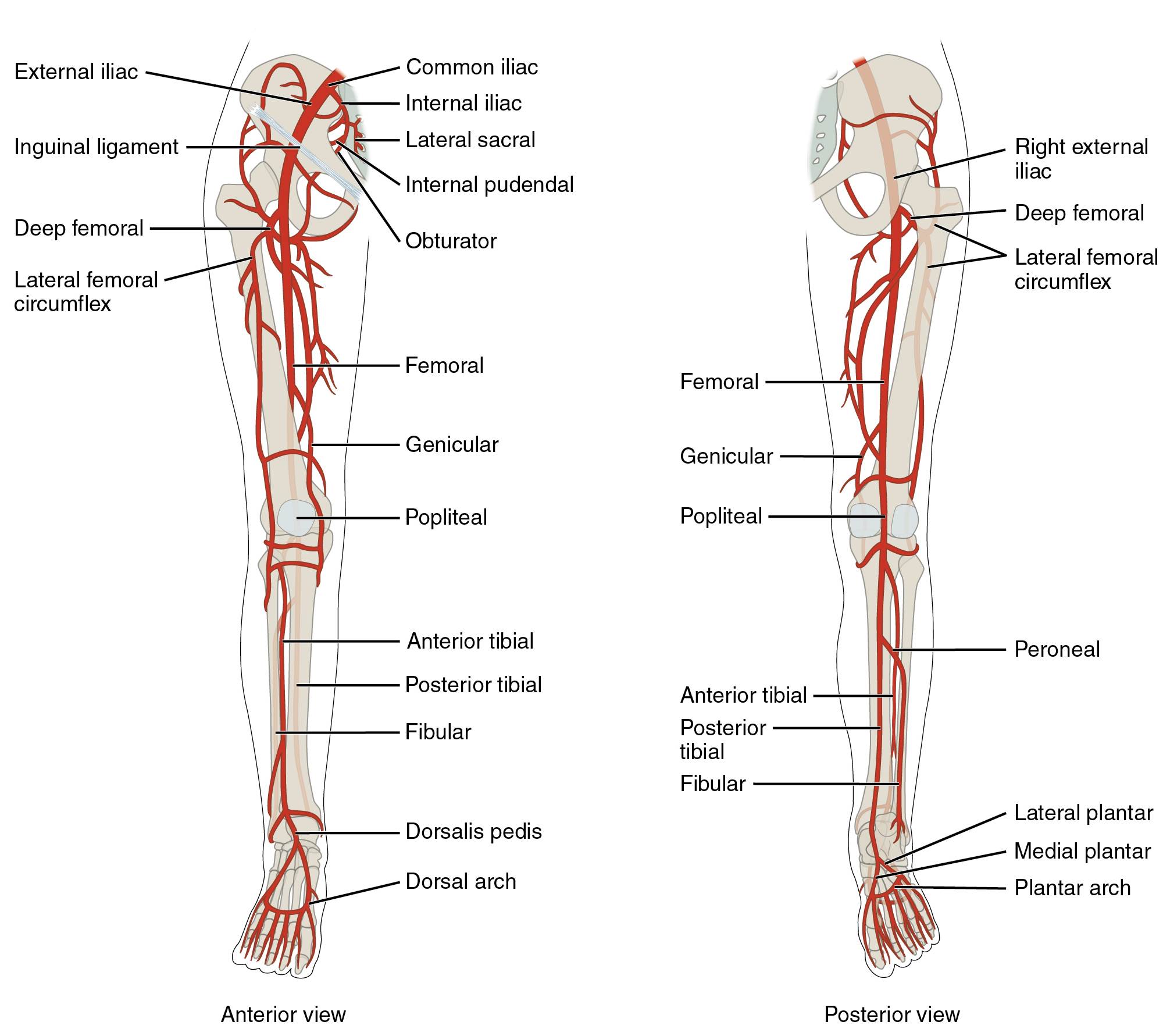
Exploring the Key Arteries of the Lower Limb
The labeled arteries in this image illustrate the primary pathways for blood supply to the lower limb, originating from the abdominal aorta. Each vessel plays a critical role in maintaining circulation to muscles, bones, and tissues.
External iliac The external iliac artery branches from the common iliac artery, passing under the inguinal ligament to become the femoral artery. It supplies blood to the pelvic region and lower limb, forming a vital link in the circulatory chain.
Common iliac The common iliac artery arises from the abdominal aorta, splitting into the internal and external iliac arteries. It serves as a primary conduit for blood flow to the pelvis and lower extremities on each side.
Internal iliac The internal iliac artery branches from the common iliac, supplying the pelvic organs, gluteal muscles, and perineum. Its extensive network supports reproductive and urinary functions alongside lower limb stability.
Lateral sacral The lateral sacral arteries branch from the internal iliac, providing blood to the sacral region and spinal nerves. They contribute to the vascular supply of the lower back and pelvic structures.
Internal pudendal The internal pudendal artery, a branch of the internal iliac, supplies the external genitalia and perineal region. It ensures adequate perfusion for reproductive and anal structures, supporting their function.
Obturator The obturator artery arises from the internal iliac, running through the obturator foramen to supply the medial thigh muscles. It plays a key role in hip joint and adductor muscle nourishment.
Inguinal ligament The inguinal ligament serves as a landmark where the external iliac artery transitions into the femoral artery. It marks the boundary between the abdominal and thigh vascular territories.
Deep femoral The deep femoral artery branches from the femoral artery, supplying the deep muscles of the thigh and hip joint. It provides a significant collateral circulation pathway, enhancing thigh perfusion.
Lateral femoral circumflex The lateral femoral circumflex artery branches from the deep femoral, wrapping around the femur to supply the lateral thigh muscles. It supports movements involving hip abduction and thigh stability.
Femoral The femoral artery, a continuation of the external iliac, runs down the anterior thigh, supplying the quadriceps and other thigh muscles. It is palpable in the groin, serving as a key site for pulse assessment.
Genicular The genicular arteries branch from the femoral and popliteal arteries, forming an anastomotic network around the knee. They ensure blood supply to the knee joint, supporting mobility and stability.
Popliteal The popliteal artery continues from the femoral artery behind the knee, supplying the knee joint and calf muscles. It bifurcates into the anterior and posterior tibial arteries, critical for lower leg circulation.
Anterior tibial The anterior tibial artery branches from the popliteal, running along the anterior leg to supply the shin muscles. It continues as the dorsalis pedis artery, feeding the dorsum of the foot.
Posterior tibial The posterior tibial artery, another popliteal branch, runs along the back of the leg, supplying the calf muscles and sole of the foot. It contributes to the plantar arch, ensuring foot perfusion.
Fibular The fibular artery, or peroneal artery, branches from the posterior tibial, supplying the lateral leg and ankle. It provides collateral circulation and supports the fibular muscles.
Dorsalis pedis The dorsalis pedis artery continues from the anterior tibial, running across the top of the foot. It supplies the dorsal foot structures and contributes to the dorsal arch.
Dorsal arch The dorsal arch is formed by the dorsalis pedis and other small arteries, supplying the toes and dorsal foot. It ensures comprehensive blood distribution to the foot’s upper surface.
Right external iliac The right external iliac artery mirrors the left, branching from the right common iliac to supply the right lower limb. It transitions into the femoral artery under the inguinal ligament.
Deep femoral circumflex The deep femoral circumflex artery branches from the deep femoral, encircling the femur to supply the thigh muscles. It enhances blood flow to the hip and thigh regions.
Peroneal The peroneal artery, synonymous with the fibular artery, supplies the lateral leg and ankle. It provides an alternative route for blood flow in case of occlusion.
Lateral plantar The lateral plantar artery branches from the posterior tibial, supplying the lateral sole of the foot. It contributes to the plantar arch, supporting the foot’s weight-bearing surface.
Medial plantar The medial plantar artery, also from the posterior tibial, supplies the medial sole of the foot. It nourishes the plantar muscles and contributes to arch stability.
Plantar arch The plantar arch is formed by the lateral and medial plantar arteries, supplying the toes and sole. It ensures robust circulation to the weight-bearing plantar surface.
The Pathway of Lower Limb Arteries
Blood flow to the lower limb begins with the abdominal aorta, progressing through a structured arterial network. This pathway supports the leg’s demanding physical role.
- The aorta bifurcates into the common iliac arteries, initiating lower limb circulation.
- The external iliac artery becomes the femoral artery, adapting to the thigh’s muscular needs.
- The popliteal artery behind the knee splits into anterior and posterior tibial branches.
- Distal arteries like the dorsalis pedis and plantar arch ensure foot and toe perfusion.
Clinical Applications of Lower Limb Arteries
Knowledge of these arteries is crucial for diagnosing vascular conditions. Pulse points and imaging rely on their anatomical positions.
- The femoral artery is a primary site for arterial catheterization and blood pressure monitoring.
- The dorsalis pedis and posterior tibial pulses assess foot perfusion in peripheral artery disease.
- Genicular arteries support knee surgery by providing collateral flow.
- Doppler ultrasound evaluates flow in the popliteal and tibial arteries for blockages.
Physiological Functions in Lower Limb Activity
These arteries adapt to physical demands, ensuring efficient oxygen delivery. Their responsiveness enhances mobility and endurance.
- The femoral and deep femoral arteries dilate during exercise, boosting thigh muscle performance.
- Plantar and dorsal arches regulate blood flow to support standing and walking.
- Hormonal changes, such as adrenaline release, influence vascular tone in the lower limb.
- Anastomoses around the knee and ankle protect against ischemic events.
In conclusion, the major arteries of the lower limb form a resilient network that supports movement and stability. Understanding their anatomy enhances clinical skills and appreciation for the body’s circulatory design.

