The circulatory system of the upper limb is a fascinating network that ensures vital blood supply from the heart to the arms and hands. This detailed flowchart illustrates the major arteries, originating from the aortic arch and branching into a complex system that supports muscle function, skin health, and more, making it an essential study for understanding human anatomy.
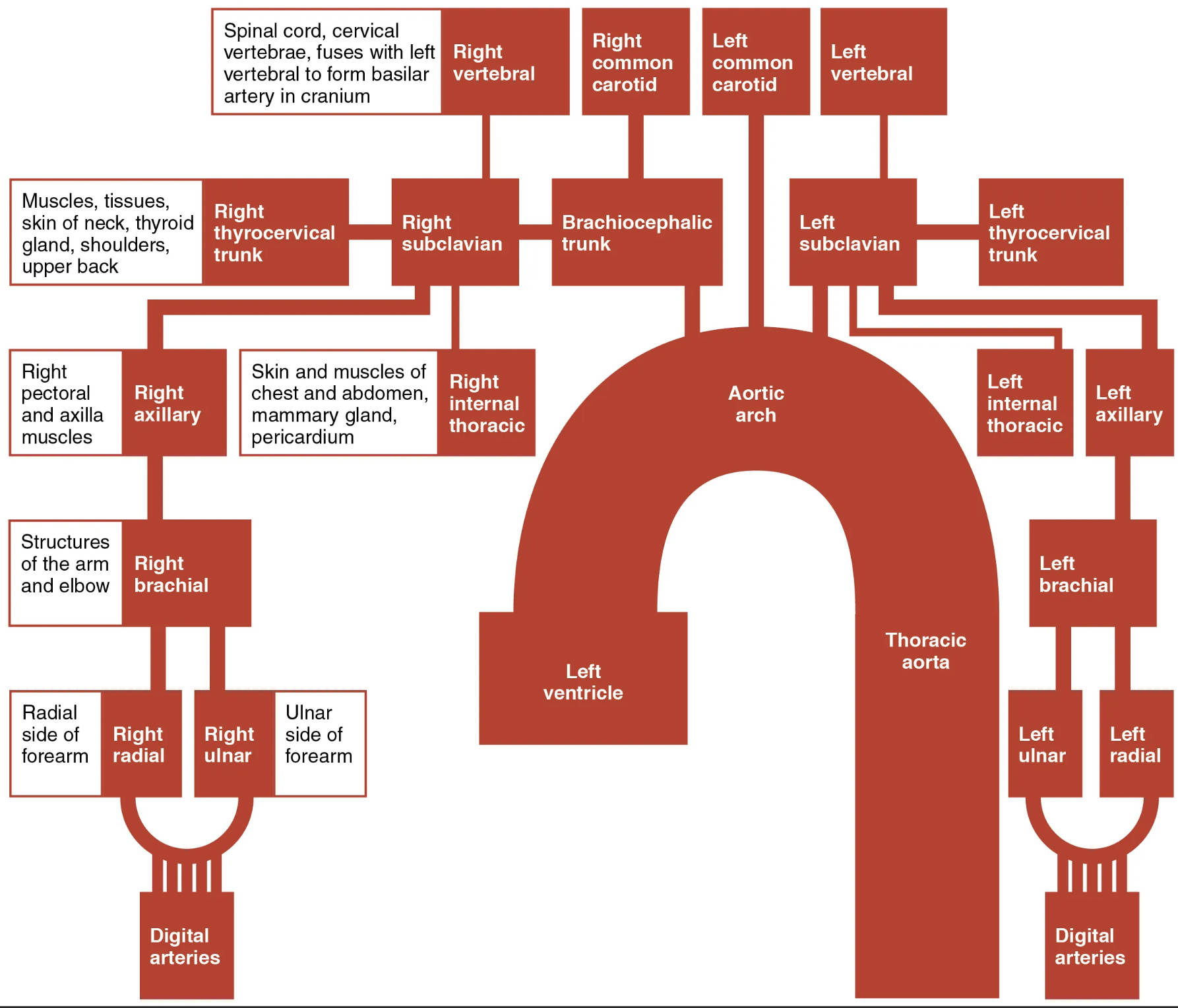
Exploring the Major Arteries in the Flowchart
The labels in this image highlight the key arteries and their roles in distributing blood throughout the upper body. Each artery contributes uniquely to the vascular network, ensuring oxygen and nutrients reach their intended destinations.
Spinal cord, cervical vertebrae, fuses with left vertebral to form basilar artery in cranium The spinal cord, cervical vertebrae, fuses with left vertebral to form basilar artery in cranium artery supplies blood to the cervical spinal cord and contributes to the basilar artery, which is critical for brain stem perfusion. This fusion enhances blood flow stability to the posterior cranial fossa, supporting vital neurological functions.
Right vertebral The right vertebral artery branches from the subclavian artery and ascends through the transverse foramina of the cervical vertebrae. It plays a key role in supplying the cerebellum and parts of the brainstem, merging with its counterpart to form the basilar artery.
Right common carotid The right common carotid artery arises from the brachiocephalic trunk, delivering blood to the head and neck, including the thyroid gland and brain. It bifurcates into internal and external carotid arteries, ensuring comprehensive cerebral circulation.
Left common carotid The left common carotid artery originates directly from the aortic arch, providing blood to the left side of the head and neck. Like its right counterpart, it splits to supply the brain and facial structures, maintaining symmetrical perfusion.
Left vertebral The left vertebral artery mirrors the right, branching from the left subclavian and ascending to join the basilar artery. It supports the posterior circulation of the brain, ensuring adequate oxygen delivery to critical areas.
Right thyrocervical trunk The right thyrocervical trunk branches from the subclavian artery, supplying the thyroid gland, neck muscles, and upper back tissues. It includes smaller arteries like the inferior thyroid, nourishing endocrine and muscular structures.
Right subclavian The right subclavian artery extends from the brachiocephalic trunk, providing blood to the right upper limb and parts of the chest. It gives rise to several branches, including the vertebral and thyrocervical arteries, before becoming the axillary artery.
Brachiocephalic trunk The brachiocephalic trunk is the first major branch off the aortic arch, splitting into the right subclavian and right common carotid arteries. It serves as a primary conduit for blood distribution to the right side of the head and arm.
Left subclavian The left subclavian artery arises directly from the aortic arch, mirroring the right subclavian’s role for the left side. It supplies the left upper limb and contributes to chest and neck circulation via its branches.
Left thyrocervical trunk The left thyrocervical trunk parallels its right counterpart, branching from the left subclavian to supply the thyroid, neck muscles, and upper back. It ensures balanced blood flow to these regions on the left side.
Skin and muscles of chest and abdomen, mammary gland, pericardium The skin and muscles of chest and abdomen, mammary gland, pericardium arteries, often internal thoracic branches, nourish the chest wall, mammary tissue, and pericardial sac. These vessels support respiratory and cardiac function through their extensive reach.
Right internal thoracic The right internal thoracic artery, a branch of the subclavian, supplies the anterior chest wall and contributes to the pericardium’s blood supply. It anastomoses with other vessels, enhancing chest wall resilience.
Aortic arch The aortic arch curves from the ascending aorta, giving rise to the brachiocephalic trunk, left common carotid, and left subclavian arteries. It serves as the central hub for systemic circulation to the upper body.
Left internal thoracic The left internal thoracic artery mirrors the right, branching from the left subclavian to supply the left chest wall and pericardium. Its anastomoses provide collateral circulation in case of blockages.
Right axillary The right axillary artery continues from the subclavian, running through the axilla to supply the shoulder and arm muscles. It transitions into the brachial artery, supporting upper limb mobility.
Right axillary muscles The right axillary muscles arteries branch from the axillary artery, nourishing the muscles of the shoulder region. These vessels ensure strength and stability during arm movements.
Structures of the arm and elbow The structures of the arm and elbow arteries, including deep brachial branches, supply the arm’s musculature and elbow joint. They maintain joint function and muscle integrity during flexion and extension.
Right brachial The right brachial artery extends from the axillary, running down the arm to supply its muscles and structures. It bifurcates into radial and ulnar arteries, critical for forearm and hand circulation.
Right ulnar side of forearm The right ulnar side of forearm artery branches from the brachial, supplying the medial forearm muscles and contributing to the palmar arches. It supports grip strength and finger movement.
Radial side of forearm The radial side of forearm artery, a brachial branch, runs along the lateral forearm to supply the thumb and lateral fingers. It is palpable at the wrist, aiding in pulse assessment.
Right radial The right radial artery continues from the radial side, forming part of the deep palmar arch in the hand. It ensures blood supply to the lateral digits, supporting fine motor skills.
Right ulnar The right ulnar artery supplies the medial forearm and contributes to the superficial palmar arch. It nourishes the ring and pinky fingers, enhancing hand functionality.
Left ventricle The left ventricle pumps oxygenated blood into the aorta, initiating the arterial flow depicted. Its powerful contractions drive circulation through the upper limb arteries.
Thoracic aorta The thoracic aorta descends from the aortic arch, supplying the chest and diaphragm via intercostal arteries. It continues as the abdominal aorta, maintaining systemic flow.
Left brachial The left brachial artery mirrors the right, supplying the left arm’s muscles and structures. It bifurcates into radial and ulnar arteries for distal circulation.
Left ulnar The left ulnar artery supplies the left medial forearm and contributes to the palmar arch. It supports the medial digits, ensuring balanced hand perfusion.
Left radial The left radial artery runs along the left lateral forearm, forming the deep palmar arch. It supplies the thumb and lateral fingers, mirroring the right side’s function.
Digital arteries The digital arteries arise from the palmar arches, running along the fingers to supply the phalanges and nail beds. They ensure fingertip sensitivity and temperature regulation.
The Origin and Pathway of Upper Limb Arteries
Blood flow begins at the heart, with the left ventricle propelling it into the aortic arch. This pathway sets the stage for a structured distribution to the upper limbs.
- The aortic arch branches into the brachiocephalic trunk, left common carotid, and left subclavian arteries.
- The brachiocephalic trunk splits to form the right subclavian and right common carotid, initiating right-side circulation.
- Subclavian arteries transition into axillary arteries, adapting to the arm’s anatomical constraints.
- Distal branching into radial and ulnar arteries ensures comprehensive hand and finger supply.
Clinical Relevance of Arterial Distribution
Understanding this arterial map aids in diagnosing circulatory issues. Accurate identification of pulse points is crucial for patient care.
- The brachial artery serves as a primary site for blood pressure measurement.
- Radial and ulnar pulses indicate distal perfusion, vital in trauma cases.
- Anomalies in vertebral artery branching can affect brain circulation, requiring imaging.
- Internal thoracic arteries support coronary artery bypass grafts, highlighting their clinical use.
Physiological Roles in Upper Limb Function
These arteries adapt to physical demands, ensuring efficient nutrient delivery. Their responsiveness enhances daily activities.
- Axillary and brachial arteries dilate during exercise, increasing muscle blood flow.
- Digital arteries regulate temperature by constricting or dilating as needed.
- Thyroid hormone influences vascular tone, impacting flow through thyrocervical branches.
- Anastomoses provide redundancy, protecting against ischemic events.
In conclusion, this flowchart offers a clear view of the arterial network sustaining the upper limb. Mastering this anatomy enhances clinical practice and deepens appreciation for the body’s circulatory efficiency.

