The lesser tubercle of the right humerus is a critical anatomical landmark in the shoulder region, playing a key role in the stability and movement of the upper arm. This article delves into the detailed anatomy of the lesser tubercle, its physical characteristics, and its clinical relevance for medical students and professionals exploring shoulder mechanics.
Labeled Anatomical Features
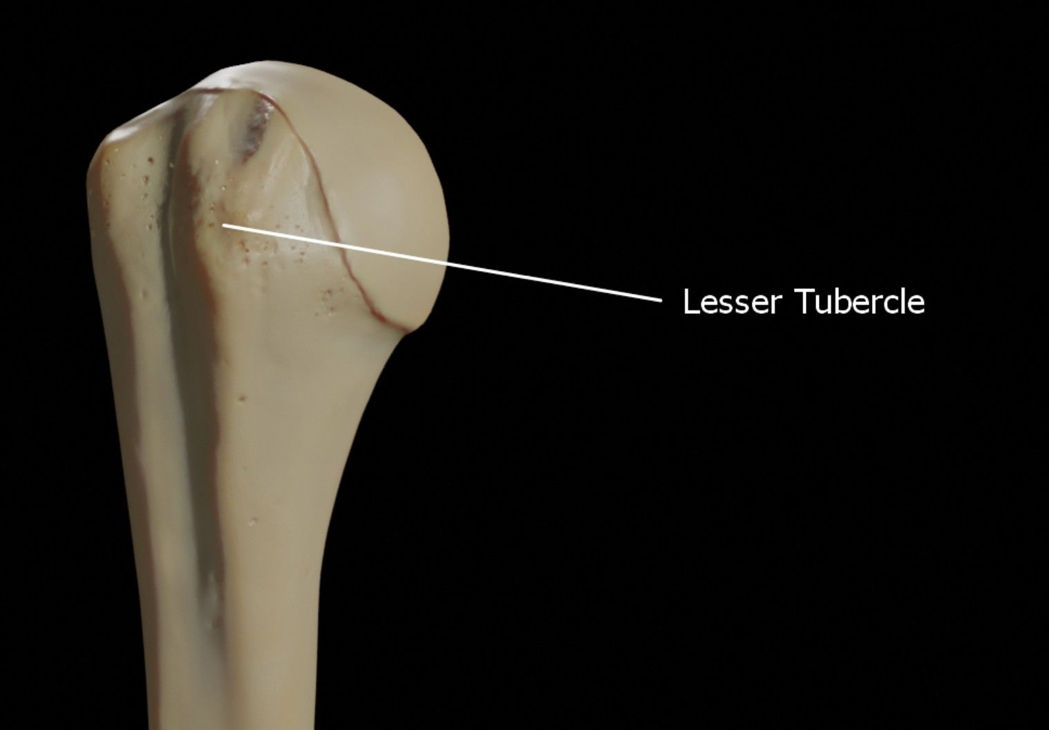
Lesser Tubercle
The lesser tubercle is a bony prominence located on the anterior aspect of the proximal humerus, just medial to the greater tubercle. It serves as the attachment site for the subscapularis muscle, a key rotator cuff muscle that facilitates internal rotation of the shoulder joint.
Detailed Anatomy of the Lesser Tubercle of the Right Humerus
Overview of the Humerus
The humerus is the long bone of the upper arm, extending from the shoulder to the elbow, and is integral to upper limb movement. Understanding its proximal features, such as the lesser tubercle, is essential for grasping shoulder joint functionality.
- The humerus consists of a proximal end, a shaft, and a distal end.
- The proximal end includes the head, anatomical neck, surgical neck, and tubercles, with the lesser tubercle being a focal point for rotator cuff muscle attachment.
- The bone is surrounded by a complex network of muscles, tendons, and ligaments that ensure stability and mobility.
Anatomical Position and Structure of the Lesser Tubercle
The lesser tubercle is a smaller, rounded projection compared to the greater tubercle, positioned on the front of the humerus. Its anatomical placement makes it a key player in shoulder joint dynamics.
- Located just below the anatomical neck of the humerus, it is separated from the greater tubercle by the intertubercular sulcus (bicipital groove).
- The lesser tubercle projects anteriorly, providing a surface for muscle attachment.
- Its smooth, rounded shape allows for efficient muscle interaction without compromising joint movement.
- The subscapularis muscle, which originates from the subscapular fossa of the scapula, inserts onto the lesser tubercle, contributing to internal rotation and stabilization of the shoulder joint.
Physical Characteristics of the Lesser Tubercle
The physical features of the lesser tubercle are tailored to its role in muscle attachment and shoulder mechanics. These characteristics are vital for medical students studying upper limb anatomy.
- The lesser tubercle is smaller than the greater tubercle, with a diameter typically ranging from 1 to 2 centimeters.
- Its surface is slightly roughened to facilitate the attachment of the subscapularis tendon.
- The bone in this region is composed of cancellous (spongy) bone internally, covered by a thin layer of compact bone, which provides strength while keeping the structure lightweight.
- The lesser tubercle’s anterior orientation ensures that the subscapularis muscle can exert force efficiently during internal rotation movements.
Functional Role in Shoulder Movement
The lesser tubercle’s primary function is to anchor the subscapularis muscle, which is crucial for shoulder joint stability and movement. This section explores its biomechanical significance.
- The subscapularis muscle, by attaching to the lesser tubercle, helps internally rotate the humerus, a motion essential for activities like reaching across the body.
- It also contributes to the stability of the glenohumeral joint by preventing anterior dislocation of the humeral head.
- During shoulder abduction, the lesser tubercle moves in coordination with the greater tubercle, ensuring smooth articulation with the glenoid cavity of the scapula.
- Its position relative to the intertubercular sulcus allows the long head of the biceps brachii tendon to glide smoothly during arm movements.
Clinical Relevance of the Lesser Tubercle
Understanding the lesser tubercle’s anatomy is crucial for diagnosing and treating shoulder-related conditions. This section highlights its clinical importance.
- Fractures involving the proximal humerus, including the lesser tubercle, can occur due to trauma, such as falls on an outstretched arm, and may disrupt subscapularis function.
- Rotator cuff injuries, particularly involving the subscapularis muscle, often affect the lesser tubercle, leading to pain and limited internal rotation.
- In shoulder dislocations, the lesser tubercle may contribute to soft tissue impingement, complicating recovery if not addressed properly.
- Surgical procedures, such as rotator cuff repairs, may involve the lesser tubercle, requiring precise knowledge of its anatomy to avoid complications.
Imaging and Diagnostic Considerations
Medical imaging, such as X-rays and MRI, plays a vital role in assessing the lesser tubercle and surrounding structures. This section discusses diagnostic approaches.
- X-rays can reveal fractures or deformities of the lesser tubercle, often taken in anteroposterior and lateral views for a comprehensive assessment.
- MRI is preferred for evaluating soft tissue injuries, such as subscapularis tendon tears, which directly involve the lesser tubercle.
- Ultrasound may be used to assess the dynamic function of the subscapularis muscle during shoulder movement.
- Proper identification of the lesser tubercle in imaging ensures accurate diagnosis and treatment planning for shoulder pathologies.
The lesser tubercle of the right humerus, though small, plays a significant role in shoulder function and stability. By understanding its anatomy, physical characteristics, and clinical relevance, medical students and professionals can better appreciate its importance in the context of upper limb mechanics and pathology. This knowledge is foundational for diagnosing and managing shoulder-related conditions effectively.

