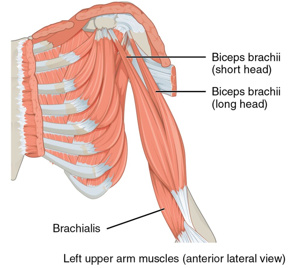
The upper arm is a vital component of the human body, housing muscles that drive essential movements of the forearm and shoulder. This article delves into the anatomy of the left upper arm muscles, as illustrated in the provided medical image, focusing on the biceps brachii (short head), biceps brachii (long head), and brachialis. These muscles play a key role in flexing, extending, pronating, and supinating the forearm, contributing to a wide range of daily activities. By examining their structure and function, readers can gain a deeper understanding of upper limb mechanics and their significance in physical health.
Introduction to the Labeled Muscles
This section offers a clear introduction to the muscles labeled in the image, providing a strong starting point. The following details explore each muscle to enhance your knowledge of upper arm anatomy.
- Biceps brachii (short head): Located on the anterior upper arm, this muscle flexes the elbow and supinates the forearm. It originates from the coracoid process of the scapula, aiding in lifting and stabilizing the arm.
- Biceps brachii (long head): Positioned alongside the short head, it also flexes the elbow and supinates the forearm. It originates from the supraglenoid tubercle, contributing to shoulder and elbow movement.
- Brachialis: Found deep to the biceps on the anterior upper arm, this muscle primarily flexes the elbow joint. It originates from the anterior humerus, providing strong elbow flexion support.
Detailed Anatomical Overview
This part provides an in-depth look at the structural details of these muscles, offering a comprehensive perspective. The biceps brachii (short head), biceps brachii (long head), and brachialis are central to upper arm function.
- The biceps brachii (short head) inserts into the radial tuberosity, innervated by the musculocutaneous nerve. Its robust structure supports powerful forearm supination and elbow flexion.
- The biceps brachii (long head) also inserts into the radial tuberosity, sharing the same innervation. Its long tendon runs through the shoulder joint, adding stability and aiding in arm elevation.
- The brachialis inserts into the ulnar tuberosity, innervated by the musculocutaneous nerve with some radial nerve contribution. Its deep position ensures consistent elbow flexion strength.
Functional Roles in the Body
This section highlights the practical applications of these muscles, emphasizing their importance in movement. The biceps brachii (short head), biceps brachii (long head), and brachialis are essential for upper limb activities.
- The biceps brachii (short head) powers lifting actions like curling weights, enhancing forearm supination. Its coordination with the long head boosts overall arm strength.
- The biceps brachii (long head) supports shoulder flexion and elbow bending, crucial for pulling motions. It stabilizes the shoulder joint during overhead activities.
- The brachialis drives elbow flexion during pulling tasks, such as rowing. Its strength complements the biceps, ensuring efficient arm movement.
Clinical Significance and Considerations
This part explores the medical relevance of these muscles, providing insights into potential health issues. The biceps brachii (short head), biceps brachii (long head), and brachialis are subject to specific conditions.
- The biceps brachii (short head) can experience strains from overuse, causing discomfort. Rest and targeted exercises help restore function.
- The biceps brachii (long head) is prone to tendonitis or ruptures, leading to pain and weakness. Physical therapy or surgery may be required for severe cases.
- The brachialis may develop myositis or strains, affecting elbow flexion. Rehabilitation focuses on strengthening and reducing inflammation.
As we conclude this examination, the biceps brachii (short head), biceps brachii (long head), and brachialis emerge as cornerstone muscles of the upper arm. Their coordinated efforts enable a range of motions from lifting to pulling, while their anatomical design supports shoulder and elbow stability. This understanding not only enriches knowledge of human anatomy but also informs effective strategies for managing related injuries, encouraging further study of their nerve supply and vascular connections.

