The human upper arm is a complex structure housing muscles critical for forearm and hand movements. This article explores the anatomy of the left upper arm muscles, showcasing their roles in flexion, extension, pronation, and supination from both anterior and posterior perspectives. The provided image highlights key muscles, offering a detailed look at their origins, insertions, and functions, essential for understanding arm mechanics and potential therapeutic interventions.
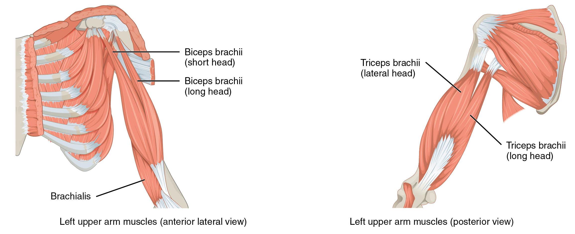
Delving into the anatomy of the upper arm provides valuable insights into its functionality. The image presents a detailed illustration of the left upper arm muscles from both anterior and posterior views, serving as an educational tool for grasping muscle distribution and action.
- Biceps brachii (short head): This muscle originates from the coracoid process of the scapula, aiding in forearm flexion and supination.
- Biceps brachii (long head): Originating from the supraglenoid tubercle, it contributes to shoulder stability and forearm flexion.
- Brachialis: Arising from the humerus, this muscle is a primary flexor of the elbow joint.
- Triceps brachii (lateral head): Originating from the humerus, it extends the forearm and stabilizes the elbow.
- Triceps brachii (long head): Stemming from the infraglenoid tubercle, it assists in arm extension and shoulder adduction.
Anatomical Overview
Exploring the muscular structure of the upper arm reveals its intricate design. The anterior view showcases muscles like the biceps brachii and brachialis, which are pivotal for elbow flexion. The posterior view highlights the triceps brachii, essential for elbow extension, demonstrating a balanced muscular framework.
- The biceps brachii, with its two heads, plays a dual role in flexing the elbow and supinating the forearm.
- The brachialis lies beneath the biceps, providing significant elbow flexion power without supination.
- The triceps brachii, comprising three heads, is the primary extensor of the forearm, with the long head also aiding shoulder movement.
Functional Roles of Upper Arm Muscles
Understanding the functional aspects enhances appreciation of arm movements. These muscles work in concert to execute precise motions, from lifting to pushing, relying on their strategic attachments.
- The biceps brachii facilitates lifting actions by flexing the elbow, crucial for activities like pulling.
- The brachialis supports continuous flexion, complementing the biceps during intense efforts.
- The triceps brachii enables pushing movements, extending the elbow for actions like pressing.
Clinical Significance
Investigating the clinical relevance underscores the importance of these muscles. Injuries or imbalances can affect daily activities, necessitating targeted rehabilitation strategies.
- Strain or tears in the biceps brachii can impair lifting capacity, often requiring physical therapy.
- Brachialis injuries may lead to weakened flexion, impacting routine tasks.
- Damage to the triceps brachii can hinder extension, potentially causing elbow instability.
Conclusion
The exploration of left upper arm muscles reveals a sophisticated interplay of anatomy and function. From the anterior biceps brachii and brachialis to the posterior triceps brachii, each muscle contributes uniquely to arm movement. Understanding these structures enhances appreciation of their roles in health and informs effective treatment approaches for related conditions, making this knowledge invaluable for practical application.

