The forearm is a dynamic region of the upper limb, housing a variety of superficial muscles that play essential roles in wrist, hand, and finger movements. This article delves into the anatomy of the left forearm superficial muscles as depicted in a palmar view, highlighting their origins, functions, and clinical relevance. The detailed illustration serves as a valuable resource for understanding the intricate muscular framework that supports everyday activities and potential therapeutic needs.
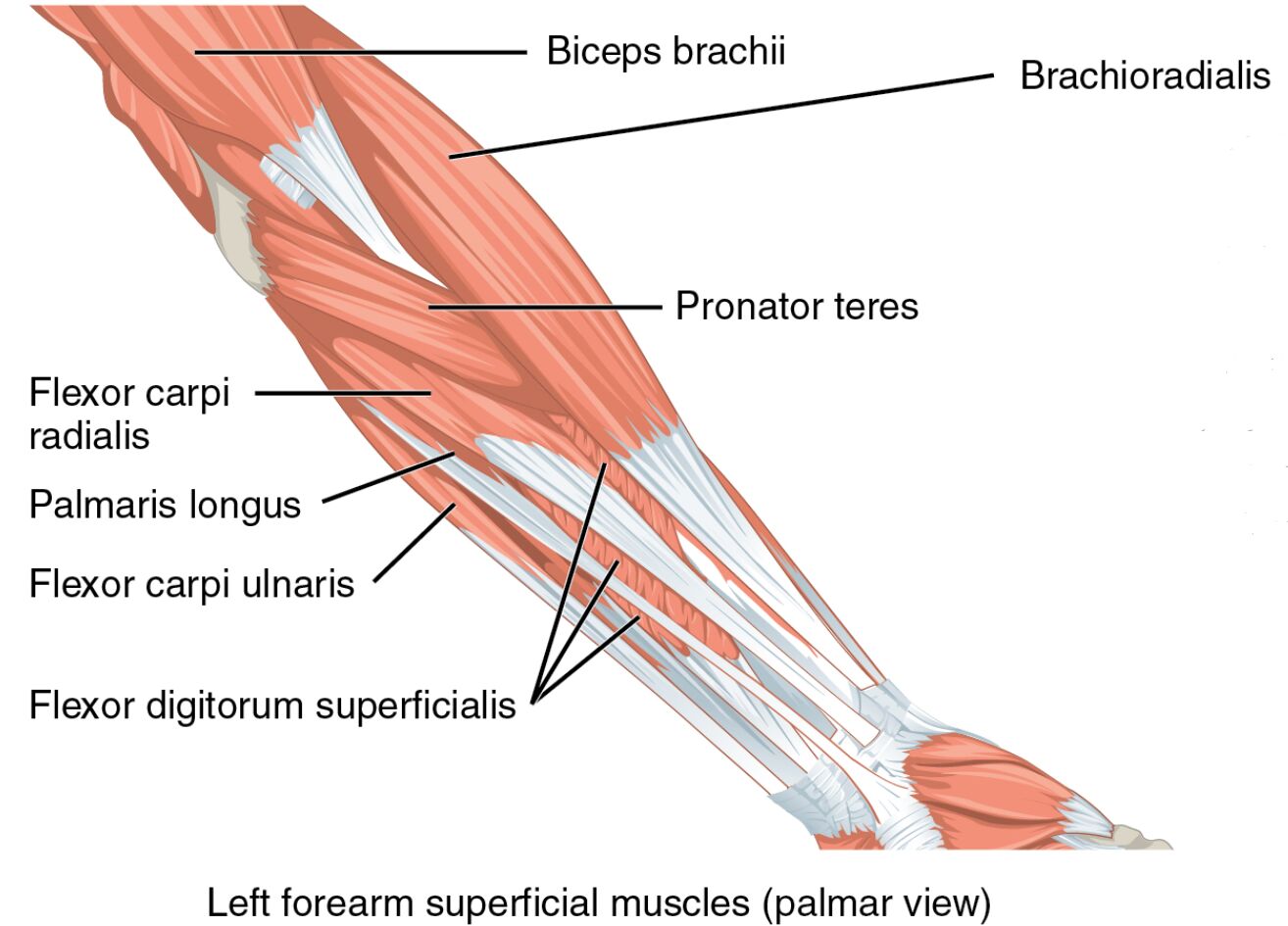
Unveiling the structure of the forearm offers a deeper understanding of its capabilities. The image provides a clear palmar view of the left forearm superficial muscles, showcasing their arrangement and labeling key components.
- Biceps brachii: Originating from the scapula, this muscle extends into the forearm, assisting in elbow flexion and forearm supination.
- Brachioradialis: Arising from the humerus, it flexes the elbow, particularly during rapid movements or with the forearm in a neutral position.
- Pronator teres: Originating from the medial epicondyle, it pronates the forearm and assists in elbow flexion.
- Flexor carpi radialis: Stemming from the medial epicondyle, it flexes and abducts the wrist, contributing to hand stability.
- Palmaris longus: Arising from the medial epicondyle, it assists in wrist flexion and tightens the palmar aponeurosis.
- Flexor carpi ulnaris: Originating from the medial epicondyle and ulna, it flexes and adducts the wrist for coordinated hand movements.
- Flexor digitorum superficialis: Arising from the medial epicondyle and radius, it flexes the middle phalanges of the fingers.
Anatomical Overview
Delving into the forearm’s muscular layout reveals its complexity. The biceps brachii and brachioradialis extend from the upper arm, while the pronator teres, flexor carpi radialis, palmaris longus, flexor carpi ulnaris, and flexor digitorum superficialis originate in the forearm, each contributing to distinct motions.
- The biceps brachii transitions into the forearm, supporting elbow flexion with its dual heads.
- The brachioradialis acts as a secondary flexor, particularly effective in neutral forearm positions.
- The pronator teres enables forearm pronation, rotating the palm downward.
- The flexor carpi radialis facilitates wrist flexion and radial deviation, aiding in hand positioning.
- The palmaris longus, when present, enhances wrist flexion and palmar support.
- The flexor carpi ulnaris drives wrist flexion and ulnar deviation, stabilizing the hand.
- The flexor digitorum superficialis allows finger flexion at the proximal interphalangeal joints.
Functional Roles of Forearm Muscles
Examining the functional aspects highlights their importance in daily tasks. These muscles work synergistically to execute precise movements, from gripping to rotating, relying on their specific attachments and innervations.
- The biceps brachii aids in lifting and supination, crucial for activities like turning a doorknob.
- The brachioradialis supports elbow flexion during rapid or resisted motions.
- The pronator teres is key for pronation, essential in tasks like using a screwdriver.
- The flexor carpi radialis contributes to wrist stability during writing or lifting.
- The palmaris longus assists in wrist flexion, often used in gripping actions.
- The flexor carpi ulnaris ensures ulnar deviation, important for fine hand adjustments.
- The flexor digitorum superficialis enables finger flexion, vital for grasping objects.
Clinical Significance
Investigating the clinical implications underscores their practical importance. Injuries or dysfunctions in these muscles can disrupt hand and wrist function, necessitating targeted interventions.
- Strain in the biceps brachii can limit supination and flexion, often treated with rest and physiotherapy.
- The brachioradialis may be affected by overuse, leading to lateral epicondylitis if untreated.
- The pronator teres, when strained, can cause pronation weakness, impacting rotational tasks.
- The flexor carpi radialis injury may result in wrist instability, requiring strengthening exercises.
- Absence or injury of the palmaris longus, though variable, can affect grip strength.
- The flexor carpi ulnaris dysfunction may lead to ulnar deviation issues, needing rehabilitation.
- The flexor digitorum superficialis, if damaged, can impair finger flexion, affecting dexterity.
Conclusion
The study of left forearm superficial muscles reveals a remarkable network of anatomy and function. From the extending biceps brachii and brachioradialis to the specialized pronator teres, flexor carpi radialis, palmaris longus, flexor carpi ulnaris, and flexor digitorum superficialis, each muscle plays a unique role in wrist and hand movements. This understanding not only enhances appreciation of the forearm’s capabilities but also informs effective strategies for managing related injuries, making it a critical area of focus for improving overall limb health.

