The forearm is a crucial part of the upper limb, featuring a rich array of superficial muscles that facilitate a wide range of movements in the wrists, hands, and fingers. This article provides an in-depth look at the left forearm superficial muscles from both palmar and dorsal perspectives, as illustrated in the accompanying image, emphasizing their anatomical structure and functional roles. This comprehensive view serves as an invaluable resource for understanding the muscular dynamics that support daily activities and inform clinical practices.
Introduction to the Image
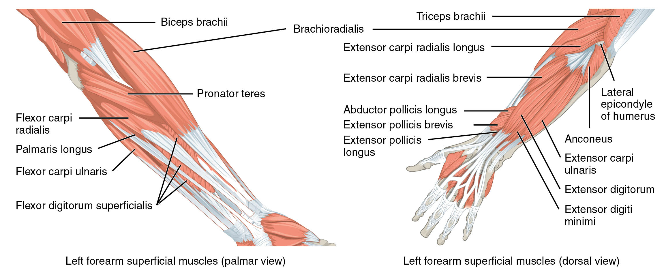
Examining the forearm from multiple angles enhances comprehension of its anatomy. The image presents the left forearm superficial muscles in both palmar and dorsal views, with detailed labels for each muscle.
- Biceps brachii: Originating from the scapula, it extends into the forearm, aiding in elbow flexion and supination.
- Brachioradialis: Arising from the lateral supracondylar ridge, it flexes the elbow, particularly in neutral positions.
- Pronator teres: Stemming from the medial epicondyle, it pronates the forearm and assists in elbow flexion.
- Flexor carpi radialis: Originating from the medial epicondyle, it flexes and abducts the wrist for hand stability.
- Palmaris longus: Arising from the medial epicondyle, it supports wrist flexion and tightens the palmar aponeurosis.
- Flexor carpi ulnaris: Stemming from the medial epicondyle and ulna, it flexes and adducts the wrist.
- Flexor digitorum superficialis: Originating from the medial epicondyle and radius, it flexes the middle phalanges of the fingers.
- Triceps brachii: Arising from the scapula and humerus, it extends the forearm and stabilizes the elbow.
- Extensor carpi radialis longus: Originating from the lateral epicondyle, it extends and abducts the wrist.
- Extensor carpi radialis brevis: Stemming from the lateral epicondyle, it assists in wrist extension and radial deviation.
- Abductor pollicis longus: Arising from the radius and ulna, it abducts and extends the thumb.
- Extensor pollicis brevis: Originating from the radius, it extends and abducts the proximal phalanx of the thumb.
- Extensor pollicis longus: Stemming from the ulna, it extends the distal phalanx of the thumb.
- Anconeus: Arising from the lateral epicondyle, it aids in elbow extension and forearm rotation.
- Extensor carpi ulnaris: Originating from the lateral epicondyle and ulna, it extends and adducts the wrist.
- Extensor digitorum: Stemming from the lateral epicondyle, it extends the fingers at the metacarpophalangeal joints.
- Extensor digiti minimi: Arising from the lateral epicondyle, it extends the little finger.
- Lateral epicondyle of humerus: A bony landmark serving as the attachment point for several forearm muscles.
Anatomical Overview
Delving into the forearm’s muscular layout reveals its dual-sided complexity. The palmar view highlights the biceps brachii, brachioradialis, pronator teres, flexor carpi radialis, palmaris longus, flexor carpi ulnaris, and flexor digitorum superficialis, which originate in the forearm and drive flexion and pronation. The dorsal view features the triceps brachii, extensor carpi radialis longus, extensor carpi radialis brevis, abductor pollicis longus, extensor pollicis brevis, extensor pollicis longus, anconeus, extensor carpi ulnaris, extensor digitorum, extensor digiti minimi, and the lateral epicondyle of humerus, focusing on extension and abduction.
- The biceps brachii and triceps brachii coordinate to balance elbow movement from opposite sides.
- The brachioradialis and pronator teres support flexion and pronation on the palmar side.
- The flexor carpi radialis and ulnaris manage wrist flexion and deviation on the palmar surface.
- The palmaris longus and flexor digitorum superficialis enhance grip and finger flexion.
- The extensor carpi radialis longus and brevis, along with the ulnaris, extend and stabilize the wrist dorsally.
- The abductor and extensor pollicis muscles provide thumb mobility and strength.
- The anconeus and extensor digitorum contribute to elbow and finger extension.
- The extensor digiti minimi specializes in little finger movement.
- The lateral epicondyle of humerus serves as a critical attachment site for dorsal muscles.
Functional Roles of Forearm Muscles
Understanding the functional dynamics underscores their importance in movement. These muscles work in harmony to execute precise actions, from flexing the wrist to extending the fingers, relying on their specific origins and insertions.
- The biceps brachii facilitates elbow flexion and supination, essential for lifting and turning.
- The brachioradialis supports rapid flexion, particularly in neutral forearm positions.
- The pronator teres enables forearm pronation, key for rotational tasks.
- The flexor carpi radialis and ulnaris drive wrist flexion and deviation, aiding hand positioning.
- The palmaris longus enhances grip strength through wrist flexion.
- The flexor digitorum superficialis allows middle finger joint flexion for grasping.
- The triceps brachii powers elbow extension, crucial for pushing actions.
- The extensor carpi radialis longus and brevis extend the wrist, supporting lifting tasks.
- The abductor pollicis longus and extensor pollicis muscles provide thumb agility.
- The anconeus assists in elbow stability during rotation.
- The extensor carpi ulnaris facilitates wrist adduction for fine adjustments.
- The extensor digitorum and digiti minimi extend the fingers, vital for hand opening.
Clinical Significance
Investigating the clinical implications highlights their practical value. Injuries or dysfunctions in these muscles can disrupt forearm and hand function, requiring tailored rehabilitation strategies.
- Strain in the biceps brachii or triceps brachii can limit elbow movement, often treated with therapy.
- The brachioradialis may develop overuse injuries, potentially leading to lateral epicondylitis.
- The pronator teres strain can impair pronation, affecting rotational ability.
- The flexor carpi radialis and ulnaris injuries may cause wrist instability, needing exercises.
- The palmaris longus absence or injury can weaken grip, though it’s variable in presence.
- The flexor digitorum superficialis damage can hinder finger flexion, impacting dexterity.
- The extensor carpi radialis longus and brevis strains may result in wrist pain, managed with rest.
- The abductor and extensor pollicis muscle injuries can reduce thumb function, requiring therapy.
- The anconeus dysfunction may lead to elbow discomfort, treatable with strengthening.
- The extensor carpi ulnaris strain can cause ulnar deviation issues, needing immobilization.
- The extensor digitorum and digiti minimi injuries can impair finger extension, necessitating care.
Conclusion
The exploration of left forearm superficial muscles from palmar and dorsal views reveals a sophisticated interplay of anatomy and function. The biceps brachii, brachioradialis, pronator teres, flexor carpi radialis, palmaris longus, flexor carpi ulnaris, flexor digitorum superficialis, triceps brachii, extensor carpi radialis longus, extensor carpi radialis brevis, abductor pollicis longus, extensor pollicis brevis, extensor pollicis longus, anconeus, extensor carpi ulnaris, extensor digitorum, extensor digiti minimi, and lateral epicondyle of humerus each contribute uniquely to forearm, wrist, and hand movements. This understanding not only enriches knowledge of muscular anatomy but also supports effective management of injuries, enhancing overall limb health and functionality.

