The forearm serves as a dynamic region of the upper limb, housing deep muscles that are crucial for the intricate movements of the wrists, hands, and fingers. This article provides a comprehensive examination of the left forearm deep muscles, presented in both palmar and dorsal views through the accompanying image, highlighting their anatomical details and functional roles. This dual-perspective analysis offers valuable insights into the deeper muscular layers that support fine motor skills and guide clinical applications.
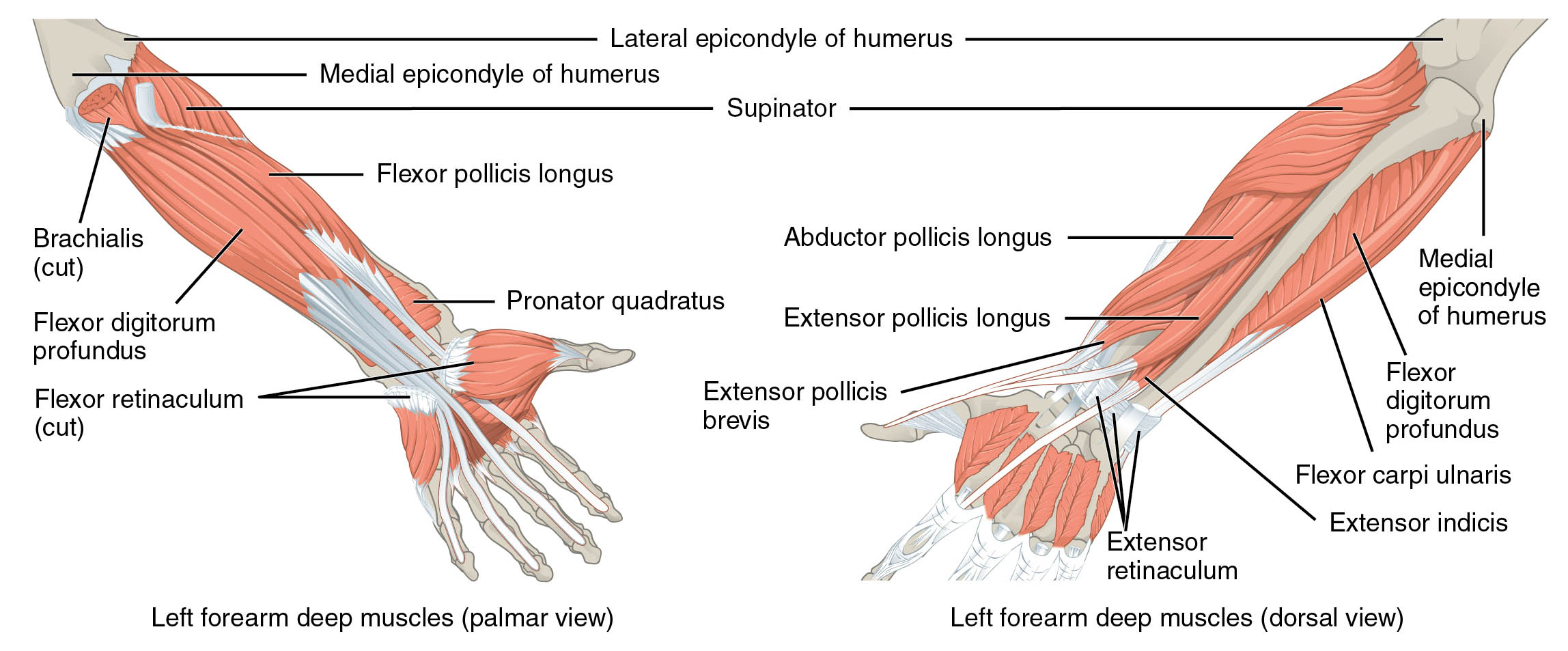
Exploring the forearm from both palmar and dorsal angles reveals its detailed structure. The image displays the left forearm deep muscles with labeled components, offering a clear view of their arrangement.
- Medial epicondyle of humerus: A bony prominence on the humerus, serving as an attachment site for flexor muscles.
- Lateral epicondyle of humerus: A humeral projection, providing anchorage for extensor muscles.
- Supinator: Originating from the lateral epicondyle and ulna, it supinates the forearm by rotating the radius.
- Flexor pollicis longus: Arising from the radius, it flexes the thumb at the interphalangeal joint.
- Brachialis (cut): Originating from the humerus, it flexes the elbow and is partially sectioned for visibility.
- Flexor digitorum profundus: Stemming from the ulna and interosseous membrane, it flexes the distal phalanges of the fingers.
- Pronator quadratus: Arising from the ulna, it pronates the forearm by rotating the radius.
- Flexor retinaculum (cut): A fibrous band, sectioned here, that stabilizes flexor tendons at the wrist.
- Abductor pollicis longus: Originating from the radius and ulna, it abducts and extends the thumb.
- Extensor pollicis longus: Stemming from the ulna, it extends the distal phalanx of the thumb.
- Extensor pollicis brevis: Arising from the radius, it extends and abducts the proximal phalanx of the thumb.
- Flexor digitorum profundus: Also visible dorsally, it flexes the distal finger phalanges from a deeper layer.
- Flexor carpi ulnaris: Originating from the medial epicondyle and ulna, it flexes and adducts the wrist.
- Extensor indicis: Stemming from the ulna, it extends the index finger for individual digit control.
- Extensor retinaculum: A fibrous band stabilizing extensor tendons at the wrist, preventing bowstringing.
Anatomical Overview
Delving into the deep muscular structure uncovers a balanced design across views. The medial epicondyle of humerus and lateral epicondyle of humerus serve as attachment points, while the supinator, flexor pollicis longus, brachialis (cut), flexor digitorum profundus, pronator quadratus, flexor retinaculum (cut) on the palmar side, and abductor pollicis longus, extensor pollicis longus, extensor pollicis brevis, flexor carpi ulnaris, extensor indicis, and extensor retinaculum on the dorsal side, drive diverse movements.
- The medial and lateral epicondyles of humerus anchor muscles for flexion and extension.
- The supinator and pronator quadratus control forearm rotation on opposite sides.
- The flexor pollicis longus and extensor pollicis muscles manage thumb flexion and extension.
- The brachialis (cut) supports elbow flexion, revealed through its sectioned view.
- The flexor digitorum profundus facilitates distal finger flexion from both perspectives.
- The flexor retinaculum (cut) and extensor retinaculum stabilize tendons on their respective sides.
- The abductor pollicis longus and extensor indicis enhance thumb and index finger mobility.
Functional Roles of Deep Forearm Muscles
Understanding the functional dynamics highlights their coordinated efforts. These muscles work together to execute precise actions, from rotating the forearm to flexing and extending digits, relying on their deep positioning.
- The supinator enables supination, essential for turning the palm upward.
- The flexor pollicis longus facilitates thumb flexion, crucial for gripping.
- The brachialis (cut) provides robust elbow flexion for lifting.
- The flexor digitorum profundus allows distal finger flexion, supporting strong grasps.
- The pronator quadratus drives pronation, rotating the palm downward.
- The flexor retinaculum (cut) maintains flexor tendon alignment at the wrist.
- The abductor pollicis longus supports thumb abduction, vital for hand function.
- The extensor pollicis longus and brevis extend the thumb, aiding precision tasks.
- The flexor carpi ulnaris contributes to wrist flexion and adduction.
- The extensor indicis extends the index finger, key for pointing.
- The extensor retinaculum stabilizes extensor tendons, ensuring smooth motion.
Clinical Significance
Investigating the clinical implications emphasizes their practical relevance. Injuries or dysfunctions in these deep muscles can disrupt hand and forearm function, requiring tailored interventions.
- Strain in the supinator can limit supination, often managed with therapy.
- The flexor pollicis longus injury may weaken thumb flexion, needing rehabilitation.
- The brachialis (cut) damage can impair elbow strength, treated with exercises.
- The flexor digitorum profundus dysfunction can affect finger flexion, impacting dexterity.
- The pronator quadratus strain may cause pronation issues, requiring strengthening.
- The flexor retinaculum (cut) compression can lead to carpal tunnel syndrome, needing surgery.
- The abductor pollicis longus injury may reduce thumb mobility, managed with therapy.
- The extensor pollicis longus and brevis dysfunction can hinder thumb extension, requiring care.
- The flexor carpi ulnaris strain may cause wrist pain, treated with rest.
- The extensor indicis injury can impair index finger movement, necessitating intervention.
- The extensor retinaculum issues, such as tendonitis, can cause wrist discomfort, treatable with support.
Conclusion
The analysis of left forearm deep muscles from palmar and dorsal views reveals a sophisticated interplay of anatomy and function. The medial epicondyle of humerus, lateral epicondyle of humerus, supinator, flexor pollicis longus, brachialis (cut), flexor digitorum profundus, pronator quadratus, flexor retinaculum (cut), abductor pollicis longus, extensor pollicis longus, extensor pollicis brevis, flexor carpi ulnaris, extensor indicis, and extensor retinaculum each contribute uniquely to forearm rotation, thumb and finger movements, and wrist stability. This dual-view understanding not only enriches knowledge of the forearm’s deeper structure but also supports effective management of injuries, enhancing overall limb health and functionality.

