The human hand is a complex and versatile structure, integral to countless daily tasks through its intricate musculature. This article examines the interossei muscles of the left hand, showcasing both palmar and dorsal views to provide a comprehensive anatomical perspective. These intrinsic muscles, which originate and insert within the hand, are crucial for fine motor control, enabling flexion, extension, abduction, and adduction of the distal finger and thumb segments. By exploring the labeled diagram, readers can gain a deeper appreciation of these muscles’ roles and their significance in hand function and clinical practice.
Introduction to the Interossei Muscles
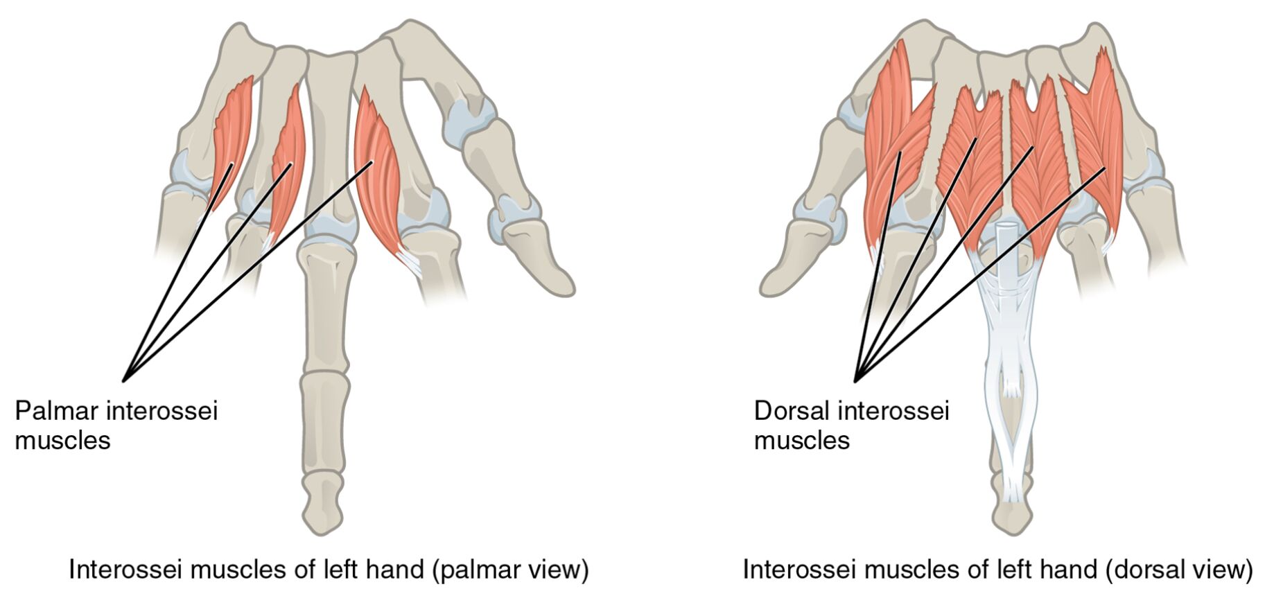
The interossei muscles are fundamental to the hand’s ability to perform precise movements. Their dual perspectives—palmar and dorsal—offer unique insights into their anatomical layout. This section delves into the labeled features that define their structure and function.
- Palmar interossei muscles: Located on the palm side, these muscles are responsible for adducting the fingers toward the middle finger. They provide essential strength for gripping and stabilizing the hand during fine motor activities.
- Dorsal interossei muscles: Positioned on the back of the hand, these muscles facilitate abduction of the fingers away from the middle finger. They are vital for actions requiring finger spreading, such as catching or balancing objects.
- Interossei muscles of left hand (palmar view): This label represents the palmar aspect of the muscle group, offering a detailed view of their arrangement. It highlights their role in enhancing hand coordination and dexterity.
- Interossei muscles of left hand (dorsal view): This label encompasses the dorsal perspective, providing a complete picture of the muscle layout. It underscores their contribution to the hand’s overall mobility and stability.
The interossei muscles‘ strategic placement enhances the hand’s versatility. Their dual views provide a holistic understanding of their anatomical and functional significance.
Detailed Anatomy of the Labeled Diagram
The image presents a side-by-side comparison of the interossei muscles from both palmar and dorsal perspectives. Each label corresponds to a specific anatomical feature, enriching the study of hand musculature.
- Palmar interossei muscles: These muscles are situated on the palm, aiding in finger adduction toward the hand’s midline. They support precision tasks like pinching or holding small objects with accuracy.
- Dorsal interossei muscles: Found on the dorsal surface, these muscles enable finger abduction away from the midline. They assist in maintaining hand balance during movements requiring finger separation.
- Interossei muscles of left hand (palmar view): This label details the palmar muscle arrangement, emphasizing their role in fine motor control. It serves as a key reference for understanding hand anatomy from this angle.
- Interossei muscles of left hand (dorsal view): This label illustrates the dorsal muscle layout, highlighting their contribution to finger spreading. It provides a critical perspective for assessing hand function.
Examining these labels reveals the interossei muscles‘ complexity. Their dual representation aids in both educational and clinical contexts, offering a complete anatomical overview.
Functional Roles of the Interossei Muscles
The interossei muscles play a pivotal role in the hand’s dynamic movements. Their functions are tailored to support a range of activities, from delicate to robust tasks.
- These muscles enable flexion and extension, allowing the fingers to bend and straighten with precision. This capability is essential for activities like writing or surgical manipulations.
- The palmar interossei muscles facilitate adduction, pulling fingers toward the midline for gripping. This action is crucial for tasks requiring a strong, controlled hold.
- The dorsal interossei muscles support abduction, spreading fingers apart for stability. This function is vital for actions like holding wide objects or performing coordinated movements.
- Their intrinsic nature ensures direct influence from the hand’s skeletal structure. This connection enhances the efficiency of finger movements in various contexts.
The interossei muscles‘ coordinated actions optimize hand performance. Their specific roles underscore their importance in maintaining dexterity and strength.
Clinical Significance and Practical Applications
The interossei muscles are frequently evaluated in clinical settings to assess hand health. Their condition directly impacts manual capabilities and quality of life.
- Injuries to the palmar interossei muscles can hinder adduction, affecting gripping strength. Rehabilitation often focuses on exercises to restore this function.
- Damage to the dorsal interossei muscles may impair abduction, limiting finger spreading ability. Targeted therapy can help regain this range of motion.
- Understanding their anatomy aids in diagnosing conditions like tendonitis or nerve damage. This knowledge guides clinicians in developing effective treatment plans.
- Their role in fine motor control makes them a focus in occupational therapy. Strengthening these muscles can improve outcomes in hand injury recovery.
This anatomical insight is invaluable for professionals. The interossei muscles‘ dual perspectives enhance diagnostic and therapeutic approaches to hand-related issues.
Conclusion
The interossei muscles of the left hand, as depicted in both palmar and dorsal views, showcase the hand’s intricate design and functionality. This article has explored their anatomical structure, diverse functional roles, and clinical relevance, providing a thorough understanding of their importance. The palmar interossei muscles and dorsal interossei muscles each contribute uniquely to hand dexterity and stability, making them essential for both everyday tasks and medical practice. A deeper study of these muscles will continue to inform therapeutic strategies and enhance appreciation of the hand’s remarkable capabilities.

