The sole of the foot contains a layered network of muscles that are crucial for supporting weight and enabling precise movements, with the intermediate layer playing a pivotal role. This article examines the intermediate muscles of the left sole, presented in a plantar view, to provide a detailed exploration of their anatomical structure and functional significance within the second layer of the plantar region. These muscles, primarily responsible for flexing and extending the toes while contributing to arch support, enhance the foot’s ability to absorb shock and maintain balance during locomotion. By analyzing the labeled diagram, readers can gain a comprehensive understanding of these muscles’ importance in foot function and their relevance in clinical practice.
Introduction to the Intermediate Muscles of the Sole
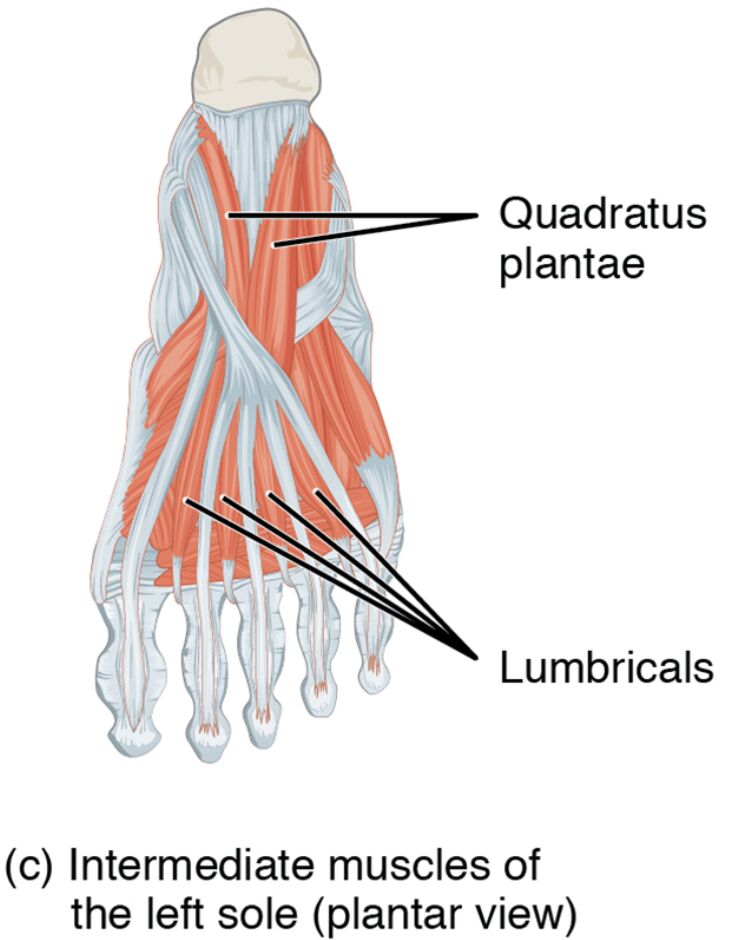
The intermediate muscles of the left sole form the second layer of the plantar surface. Their plantar view highlights their role in toe movement and foot stability. This section details the labeled structures that define their anatomy and function.
- Lumbricals: Positioned centrally in the second layer, these muscles flex the metatarsophalangeal joints and extend the interphalangeal joints. They enhance toe dexterity and assist in gripping the ground.
- Quadratus plantae (flexor accessorius): Located deeper in the second layer, it assists in flexing the toes. It supports the flexor digitorum longus in efficient toe movement.
The intermediate muscles of the left sole‘s layered placement ensures robust support. Their labeled anatomy provides a clear insight into their structural and operational roles.
Functional Roles of the Intermediate Muscles
The intermediate muscles of the left sole are essential for precise toe actions and foot support. Their positions in the second plantar layer enhance flexibility and stability. This section outlines their specific functional contributions.
- The lumbricals flex the metatarsophalangeal joints while extending the interphalangeal joints. This action improves toe control and aids in weight distribution during walking.
- The quadratus plantae assists in flexing the toes, enhancing push-off strength. It works with the flexor digitorum longus to optimize toe flexion.
The intermediate muscles of the left sole‘s coordinated efforts optimize foot performance. Their intermediate placement supports both movement and structural integrity.
Clinical Significance and Practical Applications
The intermediate muscles of the left sole are often evaluated in clinical assessments of foot health. Their condition directly impacts mobility and stability. This section explores their clinical relevance.
- Weakness in the lumbricals can lead to hammer toe or reduced toe grip. Strengthening exercises help restore toe alignment and function.
- Strain in the quadratus plantae may cause toe pain or flexor tendon issues. Stretching and conditioning alleviate discomfort and improve flexibility.
- Overuse of these muscles can contribute to metatarsalgia, affecting forefoot comfort. Rest and targeted therapy prevent further strain.
- Injury to the lumbricals may impair interphalangeal extension, impacting gait. Rehabilitation focuses on rebuilding toe strength and coordination.
- Understanding their anatomy aids in diagnosing conditions like plantar nerve entrapment. This knowledge guides effective treatment and preventive strategies.
This insight is valuable for professionals addressing foot concerns. The intermediate muscles of the left sole‘s roles underscore the need for precise therapeutic interventions.
Conclusion
The intermediate muscles of the left sole, as depicted in the plantar view, reveal the foot’s intricate muscular framework that supports toe movement and stability. This article has explored their anatomical structure, diverse functional roles, and clinical significance, providing a thorough understanding of their importance. From the lumbricals enhancing toe dexterity to the quadratus plantae aiding toe flexion, each muscle contributes uniquely to foot function and balance. Continued study of these muscles will enhance therapeutic approaches and deepen appreciation for the complex mechanics of the foot.

