The intercostal muscles are vital to the mechanics of respiration, forming layers between the ribs to support breathing and thoracic stability. This in-depth guide to the intercostal muscles anatomical structure explores their arrangement, including the external, internal, and innermost layers, providing essential insights for understanding respiratory physiology.
Labeled Parts Introduction
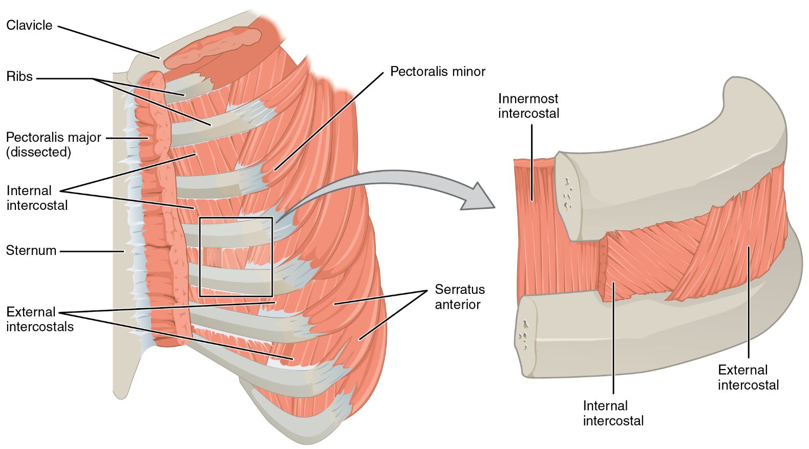
Clavicle
The clavicle is a slender bone connecting the sternum to the scapula, serving as an attachment point for muscles near the upper rib cage, and provides structural support to the shoulder. It indirectly influences the upper intercostal muscles by stabilizing the thoracic outlet.
Ribs
The ribs are curved bones forming the thoracic cage, providing attachment sites for the intercostal muscles, and protect vital organs like the heart and lungs. They move during respiration, facilitated by the contraction of the intercostal muscles.
Pectoralis major (dissected)
The pectoralis major is a large chest muscle, dissected in this image to reveal underlying structures, and assists in movements of the arm while stabilizing the rib cage. Its removal exposes the deeper intercostal layers for anatomical study.
Internal intercostal
The internal intercostal lies medially near the sternum, positioned between the external and innermost intercostal layers, and depresses the ribs during expiration to reduce thoracic volume. It plays a key role in forced exhalation and respiratory control.
Sternum
The sternum is the flat bone at the front of the chest, serving as an anterior attachment for the internal intercostal muscles, and forms part of the thoracic cage’s framework. It supports the ribs and intercostal muscles during breathing movements.
External intercostals
The external intercostals are located laterally on the sides of the body, forming the outermost intercostal layer, and elevate the ribs during inhalation to expand the thoracic cavity. They are critical for active inspiration, especially during deep breathing.
Pectoralis minor
The pectoralis minor is a smaller chest muscle beneath the pectoralis major, attaching to the ribs and scapula, and aids in stabilizing the scapula while supporting upper rib movement. It works in tandem with the intercostal muscles during respiration.
Innermost intercostal
The innermost intercostal is the deepest intercostal layer, located beneath the internal and external intercostals, and stabilizes the rib cage to prevent bulging of intercostal spaces. It assists in fine-tuning respiratory movements and supporting thoracic integrity.
Serratus anterior
The serratus anterior is a fan-shaped muscle along the lateral chest, connecting the scapula to the ribs, and protracts the scapula while aiding in rib elevation. It enhances the stability of the thoracic wall during breathing.
Overview of Intercostal Muscle Anatomy
The intercostal muscles are a series of layered muscles situated between the ribs, playing a pivotal role in respiration and thoracic cage support. This image highlights the external intercostals, internal intercostal, and innermost intercostal, with the pectoralis major (dissected) revealing their underlying structure. Their coordinated action is essential for maintaining efficient breathing mechanics.
- Forms a protective and dynamic framework around the lungs and heart.
- Facilitates the expansion and contraction of the thoracic cavity.
Structure of Intercostal Muscle Layers
The external intercostals form the superficial layer, with fibers running downward and forward, while the internal intercostal lies beneath, oriented downward and backward. The innermost intercostal, the deepest layer, runs parallel to the internal intercostals, with the sternum and clavicle providing anterior and superior attachments.
- The external intercostals are thicker and more active during inhalation.
- The internal intercostal and innermost intercostal provide stability during exhalation.
Role in Respiration
The external intercostals elevate the ribs to increase thoracic volume during inspiration, a process vital for drawing air into the lungs. The internal intercostal and innermost intercostal depress the ribs during expiration, aiding in air expulsion, while the pectoralis minor and serratus anterior support upper rib and scapular movement.
- The external intercostals enhance lung expansion during deep breaths.
- The internal intercostal assists in controlled exhalation for speech or singing.
Clinical Relevance and Physical Health
The intercostal muscles can be prone to strain, particularly the external intercostals, leading to pain during breathing. The innermost intercostal may be involved in stabilizing the rib cage after fractures, while the pectoralis minor and serratus anterior are assessed for shoulder and thoracic dysfunction.
- Weakness in the internal intercostal can impair forced exhalation.
- The serratus anterior is critical for preventing winging of the scapula.
Physical Examination and Rehabilitation
During physical exams, the clavicle and sternum are palpated to evaluate upper rib attachments, while the ribs are checked for movement range. Rehabilitation may focus on strengthening the pectoralis minor and serratus anterior to support intercostal function and improve respiratory efficiency.
- Proper alignment of the ribs ensures effective intercostal muscle action.
- The innermost intercostal is targeted in exercises to enhance thoracic stability.
Conclusion
The intercostal muscles, as illustrated in this anatomical view, form a complex system that drives respiration and maintains thoracic integrity. From the external intercostals to the innermost intercostal, with contributions from the pectoralis minor and serratus anterior, these muscles ensure efficient breathing and structural support. A deep understanding of their anatomy supports the diagnosis and treatment of respiratory and musculoskeletal conditions.

