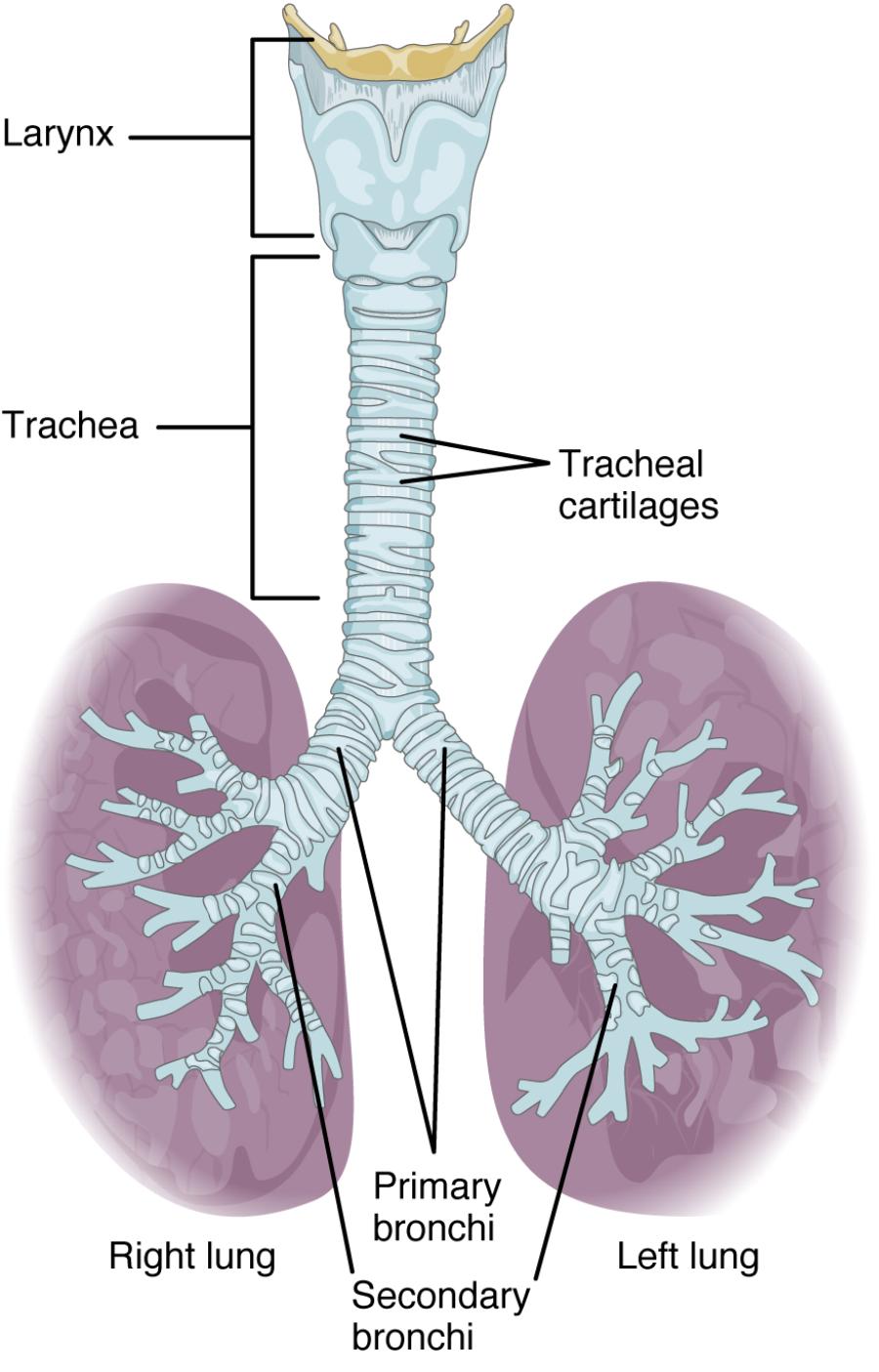
The trachea, a fundamental component of the respiratory system, serves as a conduit for air from the larynx to the lungs, supported by its distinctive C-shaped hyaline cartilage rings. This anatomical structure, formed by stacked cartilage pieces, ensures the airway remains open while allowing flexibility for adjacent structures like the esophagus. Examining this diagram provides a clear understanding of the trachea’s design and its critical role in maintaining efficient breathing.

Key Anatomical Labels in the Diagram
This section highlights each labeled part, offering a detailed explanation of their contributions to the tracheal structure.
Hyaline cartilage: Hyaline cartilage forms the C-shaped rings that provide rigid support to the trachea, preventing collapse during respiration. These rings are connected by fibrous tissue, allowing the trachea to bend slightly while maintaining an open lumen.
Tracheal lumen: The tracheal lumen is the central hollow space through which air travels to the lungs, kept patent by the surrounding cartilage. Its smooth inner lining facilitates airflow and minimizes resistance.
Smooth muscle: Smooth muscle bridges the open ends of the C-shaped cartilage rings, enabling the trachea to adjust its diameter during breathing. This muscle also contracts during coughing to expel mucus or foreign particles.
Adventitia: The adventitia is the outermost connective tissue layer, anchoring the trachea to surrounding structures like the esophagus and blood vessels. It contains nerves and vessels that supply the tracheal wall.
Submucosa: The submucosa lies beneath the mucosa, housing connective tissue, blood vessels, and glands that support the tracheal lining. It plays a role in mucus production and nutrient delivery to the epithelium.
Mucosa: The mucosa lines the inner tracheal wall, consisting of respiratory epithelium that filters and humidifies air. It contains cilia and goblet cells that work together to trap and remove debris.
Structural Design of the Trachea
The trachea’s external framework relies on its cartilage and muscle for stability. This design ensures a consistent airway under various physiological demands.
- The hyaline cartilage rings provide structural integrity, resisting compression.
- The C-shape allows esophageal expansion during swallowing without obstructing air.
- Smooth muscle adjusts the tracheal caliber, aiding in airflow regulation.
- The adventitia secures the trachea, integrating it with the neck’s anatomy.
- This configuration supports uninterrupted air delivery to the lungs.
Internal Layers and Their Functions
The tracheal wall’s internal layers enhance its protective and respiratory roles. These components work in harmony to maintain airway health.
- The mucosa filters air, trapping particles with mucus produced by goblet cells.
- Cilia within the mucosa move mucus upward, clearing the airway efficiently.
- The submucosa nourishes the epithelium, supporting its filtration function.
- Blood vessels in the submucosa deliver oxygen, sustaining ciliary activity.
- These layers prevent infections by maintaining a clean respiratory tract.
Physiological Importance in Respiration
The trachea plays a vital role in preparing air for the lungs. Its anatomy supports both structural and functional efficiency.
- The tracheal lumen ensures a clear path, humidified by mucosal secretions.
- Hyaline cartilage maintains patency, crucial during forced exhalation.
- Smooth muscle contraction aids in coughing to remove obstructions.
- The mucosa’s ciliary action forms the mucociliary escalator, protecting the lungs.
- This system optimizes gas exchange by delivering clean air.
Clinical Considerations and Anatomical Variations
Knowledge of tracheal anatomy assists in diagnosing and treating respiratory issues. Variations can impact clinical management strategies.
- Tracheal stenosis may occur if cartilage rings are malformed, narrowing the lumen.
- Excessive smooth muscle contraction can lead to airway hyperresponsiveness.
- Adventitia damage from trauma may cause tracheal displacement.
- Mucosal inflammation, as in tracheitis, can impair ciliary function.
- Imaging techniques like CT scans evaluate these structures for treatment planning.
The trachea’s ingenious design, supported by its C-shaped cartilage and layered walls, underscores its essential role in respiration. By studying this anatomical diagram, one gains a deeper appreciation for how its structure facilitates breathing, highlighting the precision and adaptability of this vital airway.

