Explore the fascinating process of how the human heart transitions from a basic structure at 28 days to a fully partitioned four-chambered organ by 8 weeks of embryonic development. This detailed guide leverages a clear diagram to highlight the anatomical changes and physiological milestones that shape the heart’s structure, offering a deep dive into its embryological evolution. From the initial separation of chambers to the formation of critical valves, this article provides a comprehensive understanding of this vital developmental phase.
Label Introduction
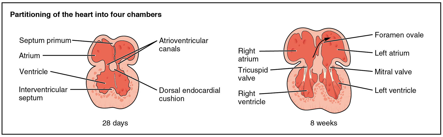
- Septum primum The septum primum is a thin wall that begins to form at 28 days, initiating the division of the atrium into left and right sides. It plays a key role in the eventual closure of the foramen ovale after birth.
- Atrium The atrium, visible at 28 days, serves as the receiving chamber for blood entering the heart, with plans to split into two distinct atria. This region is essential for coordinating blood flow into the ventricles.
- Ventricle The ventricle, present at 28 days, acts as the initial pumping chamber that will divide into left and right ventricles. Its development is crucial for the heart’s ability to pump blood effectively.
- Interventricular septum The interventricular septum starts forming at 28 days, beginning the separation of the ventricles into left and right chambers. Proper closure of this septum is vital to prevent abnormal blood mixing.
- Atrioventricular canals Atrioventricular canals, seen at 28 days, are the passages connecting the atria and ventricles, guiding early blood flow. These canals contribute to the development of the mitral and tricuspid valves.
- Dorsal endocardial cushion The dorsal endocardial cushion, forming at 28 days, is a tissue mass that helps fuse the septum primum and atrioventricular canals. It provides structural support for the heart’s chamber division.
- Right atrium The right atrium, defined by 8 weeks, receives deoxygenated blood from the body via the vena cava, marking the completion of atrial separation. It is integral to the right side of the heart’s circulatory function.
- Tricuspid valve The tricuspid valve, appearing by 8 weeks, separates the right atrium from the right ventricle, ensuring unidirectional blood flow. Its proper formation prevents backflow during ventricular contraction.
- Right ventricle The right ventricle, established by 8 weeks, pumps deoxygenated blood into the pulmonary artery for oxygenation. Its development completes the right side of the heart’s pumping system.
- Foramen ovale The foramen ovale, visible at 8 weeks, is an opening in the atrial septum allowing fetal blood to bypass the lungs. It typically closes after birth to establish adult circulation patterns.
- Left atrium The left atrium, formed by 8 weeks, receives oxygenated blood from the lungs via the pulmonary veins. It is essential for delivering oxygen-rich blood to the left ventricle.
- Mitral valve The mitral valve, present by 8 weeks, separates the left atrium from the left ventricle, ensuring efficient blood flow into the systemic circulation. Its development is critical to prevent regurgitation.
- Left ventricle The left ventricle, defined by 8 weeks, pumps oxygenated blood into the aorta for distribution to the body. Its robust development supports the high-pressure demands of systemic circulation.
Overview of Heart Partitioning
The partitioning of the heart marks a significant phase in its embryological development, starting at 28 days. This process transforms a single-chambered tube into a four-chambered organ capable of supporting complex circulation.
- Introduces the septum primum as the initial structure dividing the atrium.
- Highlights the role of the interventricular septum in separating the ventricles.
- Describes the atrioventricular canals as pathways for early blood flow.
Initial Chamber Separation at 28 Days
At 28 days, the heart begins to define its chambers with the formation of key structures. This stage lays the groundwork for the heart’s future functional divisions.
- Explains how the atrium starts receiving blood, preparing for atrial division.
- Details the ventricle‘s role as the primary pumping chamber before separation.
- Notes the contribution of the dorsal endocardial cushion to structural integrity.
Development of Atrioventricular Structures
The atrioventricular region undergoes significant changes by 28 days, shaping the heart’s internal architecture. These developments are crucial for efficient blood flow regulation.
- Describes the atrioventricular canals as connectors between atria and ventricles.
- Highlights the dorsal endocardial cushion‘s role in fusing septa and canals.
- Explains how these structures set the stage for valve formation.
Completion of Four Chambers by 8 Weeks
By 8 weeks, the heart achieves its four-chambered configuration, with distinct atria and ventricles. This milestone ensures the separation of oxygenated and deoxygenated blood.
- Details the right atrium‘s function in receiving deoxygenated blood.
- Discusses the left atrium‘s role in handling oxygenated blood from the lungs.
- Notes the right ventricle and left ventricle as specialized pumping chambers.
Role of Valves in Heart Function
Valves play a critical role in directing blood flow, with their formation completing by 8 weeks. These structures ensure the heart operates efficiently without backflow.
- Explains the tricuspid valve‘s function in the right heart, preventing regurgitation.
- Highlights the mitral valve‘s role in the left heart, supporting systemic circulation.
- Describes how these valves develop from the atrioventricular canals.
Significance of the Foramen Ovale
The foramen ovale is a unique feature of the fetal heart, facilitating circulation during development. Its presence and eventual closure are vital for postnatal adaptation.
- Details how the foramen ovale allows blood to bypass the non-functional lungs.
- Explains the septum primum‘s role in its regulation and closure after birth.
- Notes its importance in maintaining fetal oxygenation via the placenta.
Physiological and Clinical Insights
The partitioning process has profound implications for heart function and potential congenital issues. Understanding these stages aids in recognizing developmental anomalies.
- Discusses how incomplete interventricular septum closure can lead to ventricular septal defects.
- Explains how foramen ovale persistence may result in atrial septal defects.
- Highlights the role of the tricuspid valve and mitral valve in preventing circulatory inefficiencies.
Anatomical and Functional Integration
The integration of chambers and valves by 8 weeks ensures the heart can support life after birth. This final stage reflects the culmination of embryological efforts.
- Describes the right ventricle‘s adaptation for pulmonary circulation.
- Explains the left ventricle‘s development for high-pressure systemic flow.
- Notes the atrium‘s role in coordinating blood entry into the ventricles.
In conclusion, the partitioning of the human heart from 28 days to 8 weeks is a critical developmental phase that shapes its anatomical and functional capacity. The formation of the septum primum, interventricular septum, and key valves like the tricuspid valve and mitral valve ensures the heart can efficiently separate and pump blood. This diagram serves as an essential tool for understanding the heart’s evolution, offering valuable insights into both normal development and potential congenital variations.

