The lymph node serves as a critical checkpoint in the body’s immune defense, filtering lymph and activating immune responses against pathogens. Positioned along the lymphatic vessels, this small organ is a hub for lymphocyte maturation and antigen presentation, ensuring robust protection against infections. This sectional view provides a clear glimpse into its complex architecture, highlighting the interplay of various structures that sustain lymphatic function.
Labeled Components of the Lymph Node
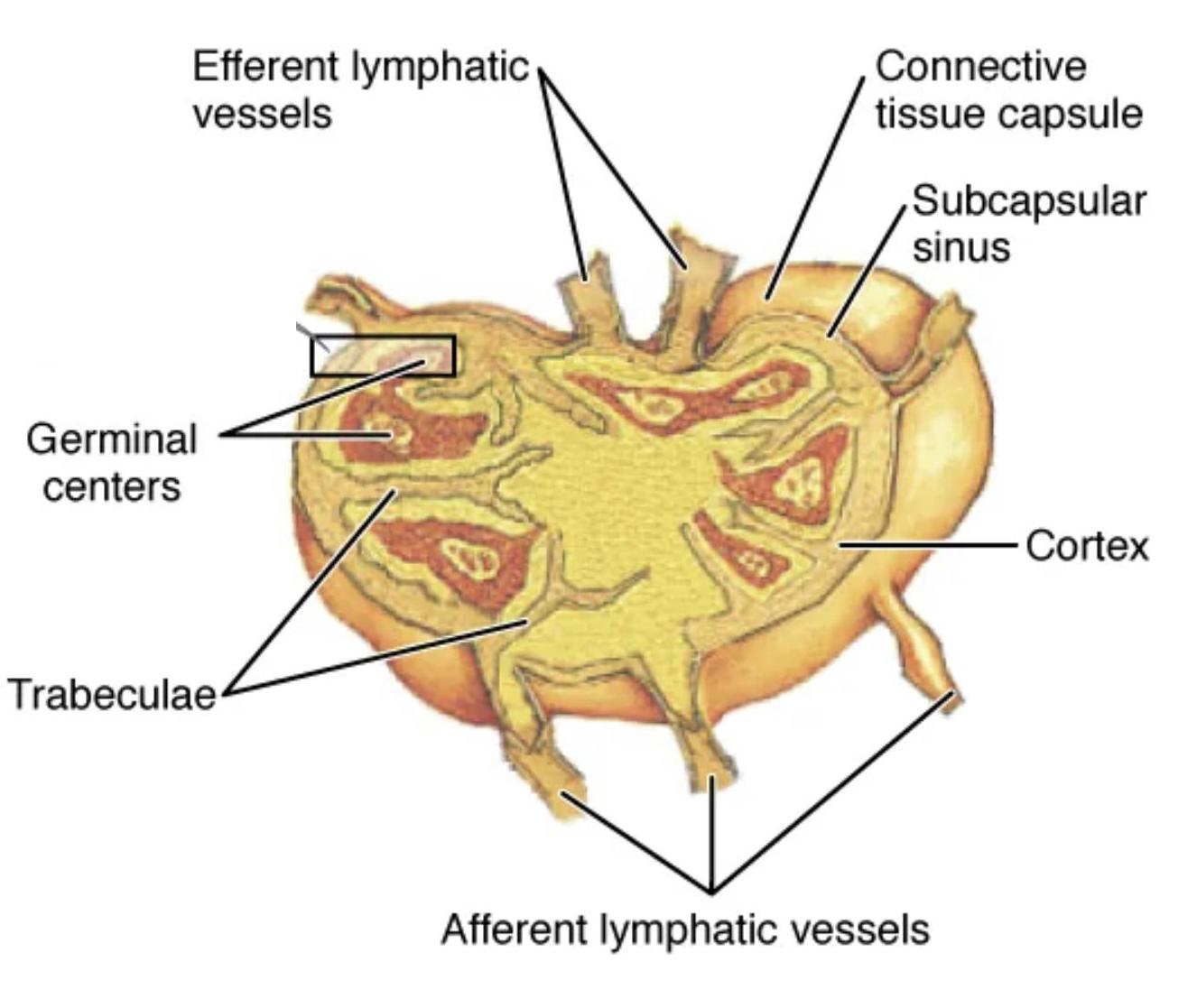
Efferent lymphatic vessels: These vessels exit the lymph node, carrying filtered lymph and activated immune cells toward the bloodstream or other nodes. They are typically located at the hilum, ensuring efficient transport of immune mediators.
Connective tissue capsule: This outer layer encases the lymph node, offering structural support and protection from external forces. It extends inward as trabeculae, dividing the node into functional compartments.
Subcapsular sinus: Positioned just beneath the connective tissue capsule, this sinus receives lymph from afferent vessels and initiates filtration. It contains macrophages that engulf pathogens, marking the first line of immune defense.
Germinal centers: Found within the cortex, these areas are sites of intense B-cell proliferation and differentiation into plasma or memory cells. They are vital for producing antibodies tailored to specific antigens.
Trabeculae: These connective tissue extensions from the capsule divide the lymph node into lobules, providing structural stability. They also guide lymph flow and support the network of blood vessels within the node.
Cortex: The outer region of the lymph node, the cortex is rich in lymphoid follicles and germinal centers where B cells are activated. It plays a central role in initiating humoral immune responses.
Afferent lymphatic vessels: These vessels bring lymph from peripheral tissues into the lymph node, delivering antigens for processing. They enter at multiple points, ensuring comprehensive filtration of lymph.
Anatomical Structure of the Lymph Node
The lymph node’s design reflects its role as an immune filter, with each component contributing to its efficiency.
- The connective tissue capsule forms a protective shell, with trabeculae extending inward to organize the internal structure.
- The subcapsular sinus serves as the entry point for lymph, where initial pathogen capture occurs.
- Germinal centers within the cortex are bustling with B-cell activity, crucial for long-term immunity.
- Afferent lymphatic vessels supply unfiltered lymph, triggering immune activation upon entry.
- Efferent lymphatic vessels exit at the hilum, exporting mature immune cells to the circulatory system.
- The cortex houses the bulk of lymphocyte activity, supported by the trabecular framework.
This sectional view emphasizes the lymph node’s layered organization, essential for its filtering role.
Physiological Role in Immune Function
The lymph node orchestrates a coordinated immune response, bridging innate and adaptive immunity.
- Lymph enters via afferent vessels, where macrophages in the subcapsular sinus begin pathogen clearance.
- In the cortex, germinal centers drive B-cell maturation, producing high-affinity antibodies.
- Trabeculae provide a scaffold, ensuring lymph flows through all regions for thorough processing.
- Efferent vessels transport activated lymphocytes, enhancing systemic immune surveillance.
- The connective tissue capsule maintains integrity, protecting against mechanical stress.
- Cytokines released within the node amplify immune cell recruitment and activity.
This dynamic process underscores the lymph node’s role in maintaining immune homeostasis.
Histological Insights and Cellular Interactions
The lymph node’s cellular landscape reveals its immunological prowess, with specialized cells working in unison.
- The cortex contains densely packed lymphocytes, with germinal centers showing mitotic B cells.
- Subcapsular sinuses host macrophages and dendritic cells, critical for antigen presentation.
- Trabeculae support reticular fibers, facilitating lymphocyte migration and lymph flow.
- Afferent vessels deliver antigens, triggering dendritic cell activation in the cortex.
- Efferent vessels carry matured immune cells, completing the immune cycle.
- The connective tissue capsule anchors blood vessels, supplying nutrients to sustain activity.
This histology highlights the node’s efficiency in immune cell education and response.
Clinical Significance of Lymph Node Anatomy
Knowledge of lymph node structure aids in diagnosing and managing immune-related conditions.
- Swollen nodes may signal infection or lymphoma, with germinal center activity as a diagnostic marker.
- The cortex’s role in B-cell maturation is key in studying autoimmune disorders.
- Blockage of afferent vessels can lead to lymphedema, emphasizing their drainage function.
- Efferent vessel dysfunction may impair immune cell dissemination, affecting recovery.
- Trabeculae and the capsule provide structural clues in chronic inflammation cases.
- Biopsies often target germinal centers to assess immune competence or malignancy.
This understanding is crucial for interpreting clinical findings and guiding treatment.
The lymph node’s intricate anatomy, as shown in this sectional view, is a testament to its vital role in immunity. By filtering lymph and activating immune cells, it ensures the body remains resilient against pathogens, making it an essential focus for those exploring human physiology and health.

