Discover the unique histological features of the large intestine, meticulously adapted for its crucial roles in water absorption, electrolyte balance, and the formation of feces. This article explores the distinctive cellular and structural components, including numerous goblet cells, deep intestinal glands, and lymphatic nodules, highlighting how these elements contribute to the large intestine’s specialized digestive functions and overall gut health.
Large intestine: This is the final segment of the gastrointestinal tract, primarily responsible for absorbing water and electrolytes, forming feces, and housing beneficial gut microbiota. Its histology reflects adaptations for these specific functions, differing significantly from the small intestine.
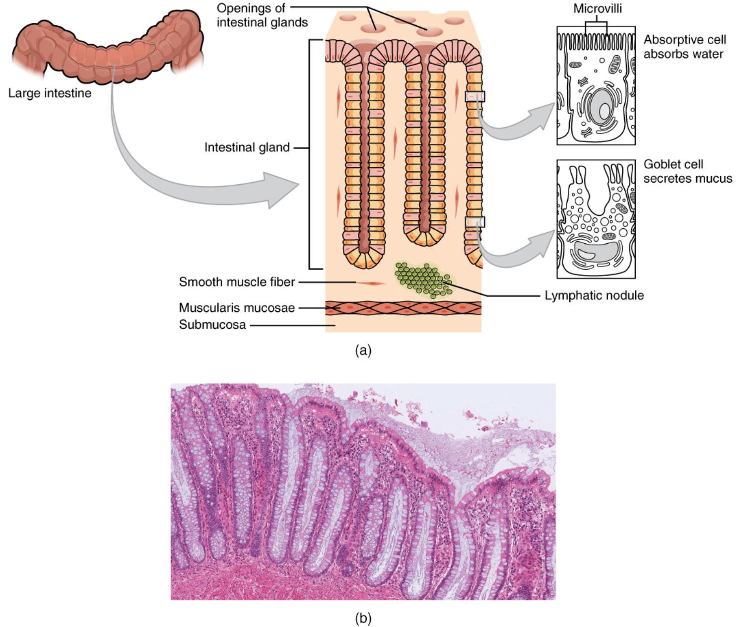
Openings of intestinal glands: These are the orifices on the luminal surface of the large intestine that lead into the crypts of Lieberkühn. These openings allow the secretions from the glandular cells to reach the lumen and facilitate the continuous renewal of the epithelial lining.
Intestinal gland: Also known as crypts of Lieberkühn, these are deep invaginations of the epithelial lining in the large intestine. They are rich in goblet cells for mucus secretion and stem cells for epithelial regeneration, crucial for maintaining the integrity of the intestinal barrier.
Microvilli: These are microscopic, finger-like projections found on the apical surface of the absorptive cells. While less prominent than in the small intestine, they still contribute to the surface area for water and electrolyte absorption.
Absorptive cell absorbs water: These columnar epithelial cells are the primary cell type in the large intestine, specialized for the reabsorption of water and electrolytes from the chyme. Their function is vital for compacting indigestible material into solid feces.
Goblet cell secretes mucus: These abundant glandular cells are interspersed throughout the epithelium of the large intestine. They produce and secrete large quantities of mucus, which lubricates the passage of feces and protects the intestinal lining from abrasion and chemical irritation.
Smooth muscle fiber: These individual muscle cells contribute to the muscular layers of the large intestine, particularly within the muscularis mucosae and muscularis externa. Their contractions facilitate segmentation and peristalsis, moving fecal matter.
Lymphatic nodule: These are clusters of lymphoid tissue located within the submucosa and lamina propria of the large intestine. They play a critical role in the immune surveillance of the gut, housing immune cells that protect against pathogens entering through the intestinal lumen.
Muscularis mucosae: This thin layer of smooth muscle is found at the base of the mucosa, underlying the intestinal glands. Its contractions contribute to local movements of the mucosa, assisting in the expulsion of gland secretions and facilitating contact with luminal contents.
Submucosa: Located beneath the muscularis mucosae, this layer consists of dense irregular connective tissue. It contains larger blood vessels, lymphatic vessels, and nerves, providing support and vascular supply to the overlying mucosa.
The large intestine, though shorter and wider than the small intestine, is a critical component of the gastrointestinal tract, primarily responsible for the final stages of digestion: water and electrolyte absorption, and the formation and temporary storage of feces. Its histological architecture is distinctively adapted to these functions, differing significantly from the small intestine’s emphasis on nutrient absorption. While the small intestine boasts elaborate villi and a dense brush border for nutrient uptake, the large intestine presents a simpler, yet highly specialized, mucosal surface.
The inner lining of the large intestine is characterized by the absence of villi and the presence of deep, straight intestinal glands, also known as crypts of Lieberkühn. These crypts are densely packed with goblet cells, which are crucial for secreting the copious amounts of mucus required to lubricate the passage of desiccating fecal matter and protect the epithelial surface. Additionally, the large intestine’s histology reveals a robust immune component, with numerous lymphatic nodules embedded within its layers, reflecting its role in immune surveillance against the vast microbial population it hosts.
Understanding the microscopic structure of the large intestine is fundamental to appreciating how it efficiently processes waste and maintains the body’s fluid and electrolyte balance. This detailed cellular and tissue organization allows for the vital reabsorption of water, transforming liquid chyme into solid feces, and ensures the safe and regulated elimination of waste products. The image above provides a clear illustration of these key histological features, emphasizing the adaptations that make the large intestine uniquely suited to its digestive and protective roles.
Key Histological Features of the Large Intestine
The large intestine’s histology is optimized for its primary functions: water absorption, electrolyte reabsorption, and the formation and elimination of feces.
- Absence of Villi: Unlike the small intestine, the large intestine lacks villi. This reflects its primary role in water and electrolyte absorption rather than nutrient absorption, which largely concludes in the small intestine. The surface area is still substantial due to the presence of deep intestinal glands and microvilli on absorptive cells, but without the extensive folding seen in the small intestine.
- Deep Intestinal Glands (Crypts of Lieberkühn): The mucosa of the large intestine is characterized by numerous straight, deep intestinal glands that extend down to the muscularis mucosae. These crypts are rich in:
- Absorptive cells: These columnar cells are specialized for water and electrolyte absorption. They possess microvilli, though less prominent than in the small intestine, which still contribute to the surface area for reabsorption. Their efficiency in water recovery is paramount for preventing dehydration.
- Goblet cells: These are exceptionally numerous in the large intestine, far more so than in the small intestine. They produce and secrete a thick, alkaline mucus. This mucus serves several vital functions: lubricating the passage of increasingly solid fecal matter, protecting the epithelium from mechanical abrasion, and neutralizing any residual acids or toxins produced by gut bacteria.
- Lymphatic Nodules: The lamina propria and submucosa of the large intestine contain abundant lymphatic nodules. These lymphoid aggregates, such as those found in Peyer’s patches in the ileum, represent a critical component of the gut-associated lymphoid tissue (GALT). They house immune cells (lymphocytes, macrophages) that constantly monitor the intestinal lumen for pathogens and mount immune responses, which is particularly important given the dense bacterial population in the large intestine.
- Muscularis Mucosae and Submucosa: Beneath the epithelial lining and lamina propria, the thin layer of muscularis mucosae supports the local movements of the mucosa. The submucosa, composed of connective tissue, contains larger blood vessels, lymphatic vessels, and the submucosal nerve plexus, providing structural support and innervation.
- Smooth Muscle Layers: The muscularis externa of the large intestine is typically composed of an inner circular and an outer longitudinal layer of smooth muscle fibers. The longitudinal layer is often condensed into three distinct bands called the teniae coli, which give the large intestine its characteristic sacculated appearance (haustra). These muscle layers are responsible for the mixing (haustral churning) and propulsive movements (mass movements) that move fecal matter towards the rectum.
In conclusion, the histology of the large intestine is a masterclass in functional adaptation. Its specialized cellular composition, particularly the abundance of goblet cells and deep intestinal glands, alongside a robust lymphatic defense system, ensures efficient water and electrolyte absorption while effectively managing waste and protecting against pathogens. This intricate microscopic structure underpins the large intestine’s vital role in maintaining fluid balance, forming feces, and contributing to overall digestive health, highlighting its unique contribution to the human body’s complex physiological processes.

