The image illustrates the gross anatomy of the lungs, showcasing their external structure and key components with clarity. This visual representation highlights the division into lobes and the branching bronchial tree, providing a foundational understanding of respiratory anatomy. It serves as an essential resource for exploring how the lungs facilitate breathing and gas exchange.
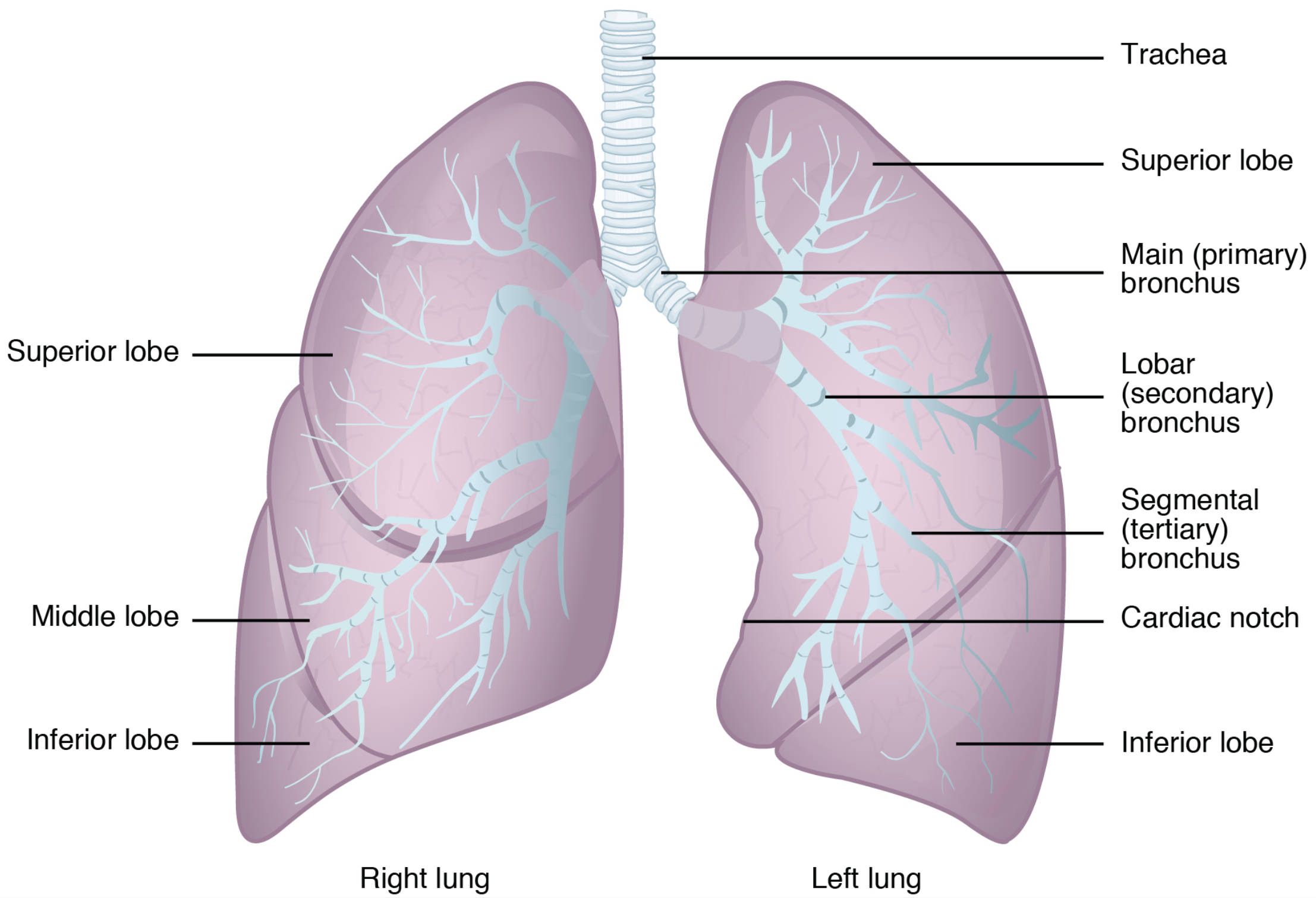
- Trachea: This rigid tube serves as the main airway, transporting air from the larynx to the bronchi, and is supported by C-shaped cartilage rings to maintain its structure. It splits into the left and right main bronchi, marking the beginning of the bronchial tree.
- Superior lobe: Located at the top of each lung, this lobe is responsible for a significant portion of gas exchange and varies slightly in size between the right and left lungs. The right superior lobe is larger, accommodating the three-lobed structure of the right lung.
- Main (primary) bronchus: Each lung receives air through a main bronchus, which branches from the trachea and is lined with cartilage to prevent collapse. The right main bronchus is shorter and more vertical, making it more prone to foreign body aspiration.
- Lobar (secondary) bronchus: These branches supply air to each lobe of the lung, further dividing the airflow to ensure even distribution. There are two lobar bronchi on the left lung and three on the right, corresponding to their respective lobes.
- Segmental (tertiary) bronchus: These smaller branches deliver air to specific segments within each lobe, enhancing the lung’s ability to ventilate different regions. They are critical for localized gas exchange and are numerous within the lung tissue.
- Cardiac notch: This indentation on the left lung accommodates the heart, allowing it to fit snugly within the thoracic cavity. It is a distinctive feature that differentiates the left lung from the right.
- Middle lobe: Present only in the right lung, this lobe lies between the superior and inferior lobes and contributes to the right lung’s three-lobed configuration. It plays a key role in the right lung’s overall respiratory function.
- Inferior lobe: The largest lobe in both lungs, it occupies the lower portion and is vital for substantial gas exchange due to its extensive surface area. It contains numerous segmental bronchi to support its size and function.
- Right lung: This lung has three lobes—superior, middle, and inferior—due to the presence of an oblique and a horizontal fissure. Its structure supports a larger capacity for air intake on the right side of the body.
- Left lung: Featuring two lobes—superior and inferior—due to a single oblique fissure, this lung is slightly smaller to accommodate the heart. Its design ensures efficient respiration despite the cardiac notch.
Anatomical Structure of the Lungs
The lungs’ gross anatomy reveals a sophisticated design tailored for respiration. Each labeled component works together to optimize oxygen intake and carbon dioxide expulsion. This structure is fundamental for understanding pulmonary function.
- The trachea acts as the central conduit, ensuring a clear path for air to reach the lungs.
- Superior lobe handles a large share of ventilation, adapting to the lung’s overall shape.
- The main (primary) bronchus divides the airflow, setting the stage for further branching.
- Lobar (secondary) bronchus ensures each lobe receives adequate air supply.
- Segmental (tertiary) bronchus fine-tunes ventilation to specific lung segments.
- The cardiac notch allows anatomical compatibility with the heart’s position.
- Middle lobe enhances the right lung’s capacity, supporting its three-lobed design.
- Inferior lobe maximizes gas exchange with its extensive alveolar network.
- Right lung’s three lobes provide a robust framework for respiration.
- Left lung adjusts its two-lobed structure to fit the thoracic cavity’s layout.
Physiological Functions of Lung Components
The physiological roles of the lung’s anatomical features are critical for sustaining life. These components enable the lungs to perform their primary role of gas exchange efficiently. Their coordinated action ensures adequate oxygenation of the blood.
- The trachea maintains airway patency, warmed and humidified air before it reaches the bronchi.
- Superior lobe contributes to upper respiratory function, adjusting to positional changes.
- The main (primary) bronchus delivers air with minimal resistance, thanks to cartilage support.
- Lobar (secondary) bronchus distributes air evenly across lobar regions.
- Segmental (tertiary) bronchus supports localized ventilation, enhancing oxygen delivery.
- The cardiac notch ensures the heart and lung coexist without compromising function.
- Middle lobe aids in right lung ventilation, complementing the inferior lobe’s role.
- Inferior lobe houses the majority of alveoli, critical for extensive gas exchange.
- Right lung’s larger volume supports greater oxygen intake on that side.
- Left lung compensates for its size with efficient two-lobed ventilation.
Clinical Relevance and Educational Insights
The gross anatomy of the lungs provides a basis for diagnosing and treating respiratory conditions. Understanding these structures aids in identifying abnormalities like pneumonia or lung cancer. This knowledge is invaluable for developing effective treatment plans.
- The trachea can be a site for tracheal stenosis if scarred, affecting airflow.
- Superior lobe is often a primary site for tuberculosis infections due to gravity.
- The main (primary) bronchus’ angle influences foreign body lodgment risks.
- Lobar (secondary) bronchus involvement can indicate lobar pneumonia.
- Segmental (tertiary) bronchus issues may lead to segmental lung collapse.
- The cardiac notch’s proximity to the heart can complicate left lung surgeries.
- Middle lobe syndrome can occur if airflow is obstructed in the right lung.
- Inferior lobe is prone to aspiration pneumonia due to its lower position.
- Right lung’s extra lobe increases its susceptibility to certain infections.
- Left lung’s cardiac notch affects its vulnerability to pressure changes.
The gross anatomy of the lungs, as depicted, offers a clear view of their structural elegance and functional importance. These components work seamlessly to support respiration, providing a solid foundation for further study and clinical application. Exploring this anatomy deepens appreciation for the lungs’ role in maintaining overall health.

