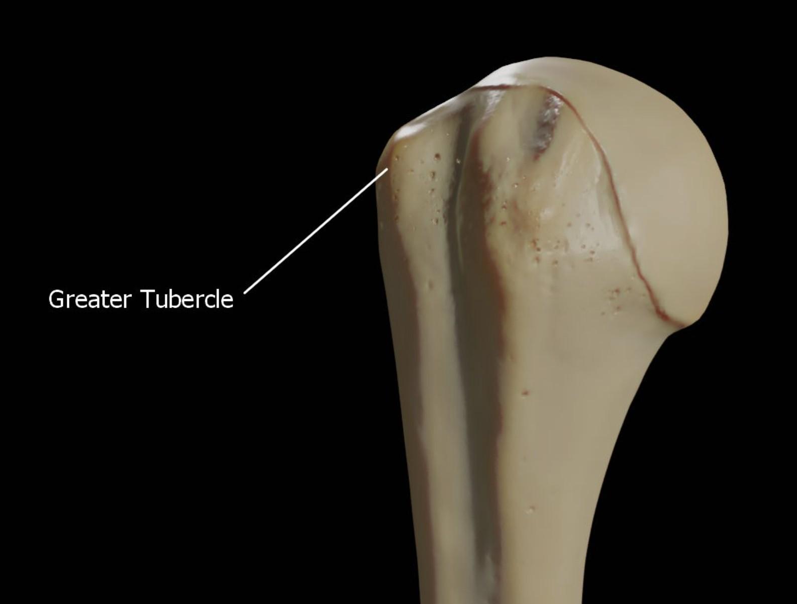
The greater tubercle of the right humerus is a critical bony landmark in the upper arm, playing a significant role in shoulder function and stability. This medical image highlights the greater tubercle, offering a clear visual for medical students and professionals studyingupper limb anatomy. In this article, we explore the labeled greater tubercle, its anatomical features, physical characteristics, and clinical relevance to provide a comprehensive understanding of its importance in the shoulder joint.
Labeled Part of the Right Humerus
This section explains the labeled structure in the image, providing a detailed description of its role in the humerus.
Greater Tubercle
The greater tubercle is a prominent bony projection on the lateral side of the proximal humerus, just below the anatomical neck. It serves as the attachment site for three rotator cuff muscles—supraspinatus, infraspinatus, and teres minor—making it essential for shoulder movement and stability.
Anatomy of the Greater Tubercle of the Right Humerus
The greater tubercle is an integral part of the proximal humerus, contributing to the shoulder’s structure and function. This section delves into its anatomical features and relationships.
- Location and Structure: The greater tubercle is situated laterally on the proximal humerus, adjacent to the lesser tubercle and below the humeral head. It has a roughened surface with three distinct facets for muscle attachments.
- Muscle Attachments: The supraspinatus attaches to the superior facet, the infraspinatus to the middle facet, and the teres minor to the inferior facet of the greater tubercle. These attachments enable abduction, external rotation, and stabilization of the shoulder joint.
- Relation to the Shoulder Joint: The greater tubercle lies just outside the anatomical neck, contributing to the shoulder joint’s stability by anchoring the rotator cuff muscles. It also forms part of the bicipital groove’s lateral border, which houses the biceps tendon.
- Neurovascular Proximity: The greater tubercle is near the axillary nerve and posterior circumflex humeral artery, which run posteriorly to the humerus. Injuries to the greater tubercle can potentially affect these structures, leading to complications.
Physical Characteristics of the Greater Tubercle
Understanding the physical properties of the greater tubercle is crucial for clinical assessment and diagnosis. This section covers its physical features and examination techniques.
- Surface Features: The greater tubercle has a rough, irregular surface due to its muscle attachment sites, making it distinguishable from the smoother humeral head. Its lateral position can be palpated beneath the deltoid muscle.
- Palpation Techniques: Clinicians can palpate the greater tubercle by pressing laterally just below the acromion, especially during shoulder abduction or external rotation. This helps assess for tenderness or deformities following trauma.
- Radiographic Appearance: On X-rays, the greater tubercle appears as a lateral prominence on the proximal humerus, often overlapping with the humeral head in certain views. Fractures or dislocations involving the greater tubercle are typically visible on anteroposterior or axillary radiographs.
- Range of Motion Impact: The greater tubercle’s role in anchoring rotator cuff muscles means it directly influences shoulder abduction and external rotation. Damage to this area can lead to impaired shoulder mobility and weakness.
Clinical Relevance of the Greater Tubercle in Shoulder Function
The greater tubercle is often involved in shoulder injuries and conditions, with significant clinical implications. This section explores common issues and their effects.
- Greater Tubercle Fractures: Fractures of the greater tubercle are common in shoulder dislocations or direct trauma, often occurring alongside anterior dislocations. These fractures can disrupt rotator cuff function, leading to weakness in abduction and external rotation.
- Rotator Cuff Injuries: Since the greater tubercle anchors the supraspinatus, infraspinatus, and teres minor, rotator cuff tears can affect its biomechanical role. Chronic tears may lead to bone remodeling or degeneration of the greater tubercle.
- Shoulder Dislocation Complications: Anterior shoulder dislocations can cause a greater tubercle fracture or a Hill-Sachs lesion on the humeral head, impacting the greater tubercle’s alignment. This may result in shoulder instability and recurrent dislocations.
- Surgical Considerations: During rotator cuff repair surgeries, the greater tubercle serves as a key landmark for reattaching the torn tendons. Surgeons often use suture anchors placed in the greater tubercle to restore rotator cuff integrity.
Functional Role of the Greater Tubercle in Shoulder Movement
The greater tubercle plays a pivotal role in facilitating shoulder movements through its muscle attachments. This section highlights its contributions to shoulder mechanics.
- Shoulder Abduction: The supraspinatus, attached to the greater tubercle, initiates shoulder abduction, allowing the arm to lift overhead. Damage to the greater tubercle can impair this motion, affecting daily activities like reaching.
- External Rotation: The infraspinatus and teres minor, both anchored to the greater tubercle, are primary external rotators of the shoulder. These muscles are crucial for movements like throwing or combing hair.
- Shoulder Joint Stability: The rotator cuff muscles attached to the greater tubercle work together to stabilize the glenohumeral joint, preventing dislocation during dynamic activities. The greater tubercle’s structural integrity is essential for this stability.
- Force Transmission: The greater tubercle acts as a lever arm for the rotator cuff muscles, transmitting forces from the muscles to the humerus. This mechanism ensures efficient shoulder movement and load distribution.
The greater tubercle of the right humerus is a vital structure for shoulder function, serving as a key attachment point for the rotator cuff muscles and contributing to shoulder stability and movement. By understanding its anatomy, physical characteristics, and clinical significance, medical students can better appreciate its role in the upper limb and its implications in shoulder-related conditions, enhancing their diagnostic and clinical skills.

