Granular leukocytes, a vital subset of white blood cells, play a crucial role in the body’s immune defense by targeting pathogens and mediating inflammatory responses. This diagram showcases the distinct appearances and functions of neutrophils, eosinophils, and basophils, highlighting their unique granular structures and nuclear shapes under a microscope. Exploring these cells provides a deeper understanding of their specialized contributions to health and immunity.
Key Components of Granular Leukocytes
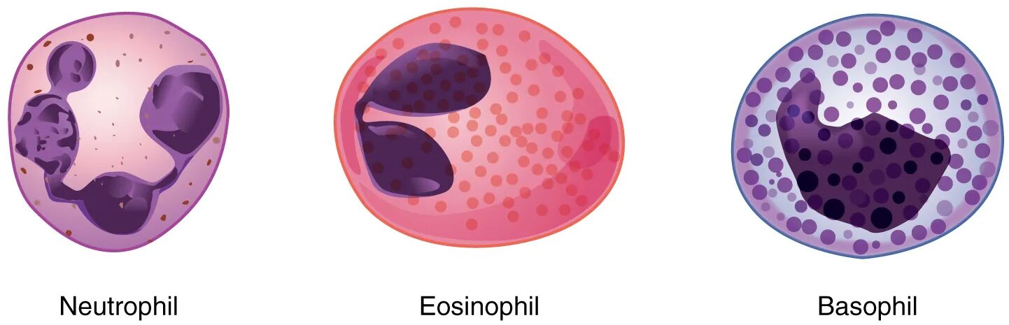
The diagram illustrates the morphological and functional characteristics of each granular leukocyte type.
Neutrophil:
The neutrophil is the most abundant granular leukocyte, featuring small, light lilac-staining granules and a nucleus with two to five lobes. It acts as a first responder in bacterial infections, rapidly phagocytizing pathogens and releasing antimicrobial substances.
Eosinophil:
The eosinophil contains slightly larger granules that stain reddish-orange, with a nucleus typically consisting of two to three lobes, aiding in its identification. It targets parasitic infections and modulates allergic responses by releasing cytotoxic proteins.
Basophil:
The basophil is characterized by large, dark blue to purple granules that often obscure its two-lobed nucleus, making it the least common granular leukocyte. It releases histamine and heparin during allergic reactions and inflammation, contributing to immediate immune responses.
The Anatomical and Physiological Role of Granular Leukocytes
Granular leukocytes are essential components of the innate immune system, each with specialized roles in protecting the body. The neutrophil, with its multi-lobed nucleus and light lilac granules, dominates acute bacterial infections, employing phagocytosis to engulf and destroy invaders, often forming pus as a byproduct. Eosinophils, with their reddish-orange granules, are particularly effective against parasites like helminths, releasing major basic protein to disrupt their membranes, and are also involved in allergic conditions by counteracting histamine effects.
Basophils, despite their scarcity, play a pivotal role in allergic and anaphylactic responses, with dark purple granules storing histamine and heparin to promote vasodilation and prevent clotting. These cells are produced in the bone marrow, regulated by cytokines like granulocyte colony-stimulating factor (G-CSF) and interleukin-5 (IL-5), which are influenced indirectly by hormones such as T3 and T4 from the thyroid gland that affect metabolic demand. Their granular contents are preformed mediators, ready for rapid release upon activation.
- Neutrophil Functions: Releases neutrophil extracellular traps (NETs) to capture bacteria; lifespan is short, lasting hours to days.
- Eosinophil Activities: Increases in number during asthma attacks; granules contain peroxidase for oxidative bursts.
- Basophil Contributions: Secretes leukotrienes to amplify allergic inflammation; rare but critical in hypersensitivity.
Physical Characteristics and Clinical Relevance
The physical traits of granular leukocytes, as depicted, are key to their identification and function under the microscope. Neutrophils exhibit a segmented nucleus and pale granules, reflecting their readiness for phagocytosis, while eosinophils’ larger, vividly stained granules signal their parasitic focus. Basophils stand out with dense, obscuring granules and a bilobed nucleus, indicating their role in immediate hypersensitivity.
Clinically, these characteristics guide diagnosis and treatment. Elevated neutrophil counts suggest bacterial infections or inflammation, often assessed via complete blood count (CBC) with differential. Increased eosinophils may indicate parasitic infections or allergies like asthma, while high basophil levels point to chronic allergic conditions or myeloproliferative disorders. Therapies include corticosteroids to reduce eosinophil activity in allergies or G-CSF to boost neutrophil production in neutropenia.
- Diagnostic Tools: Blood smears reveal granule staining; flow cytometry quantifies leukocyte subsets.
- Therapeutic Options: Antihistamines target basophil-mediated allergies; antiparasitic drugs address eosinophil-driven infections.
Conclusion
The granular leukocytes diagram offers a clear view of the diverse roles played by neutrophils, eosinophils, and basophils in immune defense, from combating bacteria to managing allergies. These cells’ distinct structures and functions highlight the body’s sophisticated response to various threats, ensuring rapid and targeted protection. By understanding their characteristics, one can better appreciate the diagnostic and therapeutic approaches that support immune health, emphasizing their importance in maintaining physiological balance.

