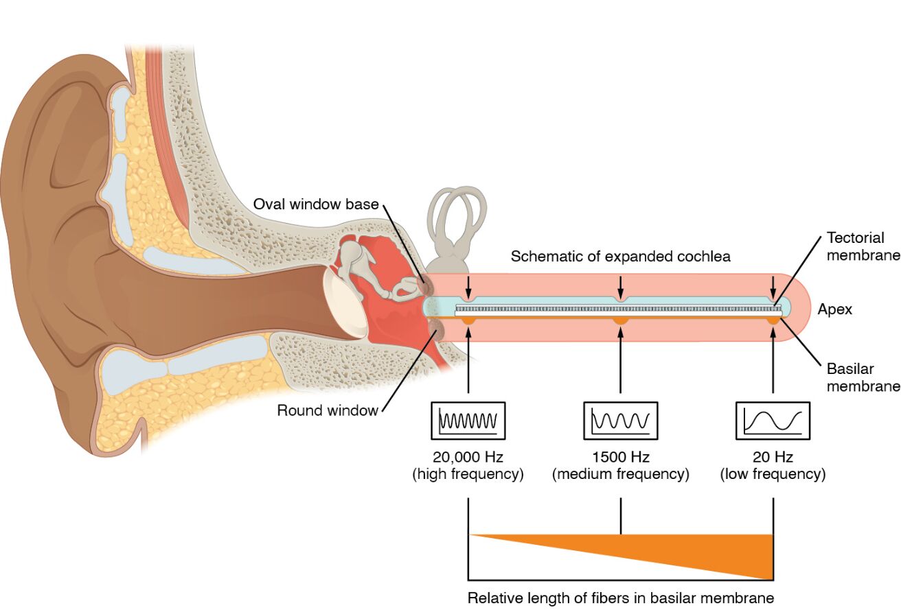
The cochlea serves as the inner ear’s masterpiece, transforming sound waves into electrical signals through a process of frequency coding that varies along its length, as depicted in this image. This image illustrates how the movement of the oval window generates a standing sound wave that deflects the basilar membrane, activating hair cells at different cochlear regions based on sound frequency—high at the base and low at the apex. This article explores the anatomical and physiological mechanisms behind this frequency coding, providing a detailed insight into how the cochlea decodes the complexity of sound.
Labeled Parts of the Cochlea
Cochlea The cochlea is a spiral, fluid-filled structure in the inner ear that houses the organ of Corti and processes sound vibrations into neural signals. Its length is divided into regions that respond to different sound frequencies, enabling detailed auditory perception.
Basilar membrane The basilar membrane supports the organ of Corti and varies in width and stiffness from the base to the apex of the cochlea, determining frequency-specific responses. Its deflection by sound waves activates hair cells, tuning the ear to different pitches.
Hair cells Hair cells are mechanoreceptor cells within the organ of Corti, with stereocilia that bend in response to basilar membrane movement to generate electrical signals. They are distributed along the cochlea, with basal cells responding to high frequencies and apical cells to low frequencies.
Oval window The oval window is a membrane-covered opening that receives amplified vibrations from the stapes, initiating pressure waves in the cochlear fluid. Its movement sets the basilar membrane into motion, driving the frequency coding process.
Apex The apex is the wide, flexible end of the cochlea, where the basilar membrane is tuned to respond to low-frequency sounds. Hair cells in this region detect deeper tones, contributing to the perception of bass sounds.
Base The base is the narrow, stiff end of the cochlea, where the basilar membrane responds to high-frequency sounds. Hair cells here are activated by higher pitches, enabling the perception of treble sounds.
Anatomical Overview of Frequency Coding
The cochlea’s structure is designed to encode sound frequency through the differential response of its basilar membrane and hair cells. This anatomical gradient allows for precise auditory discrimination across a wide range of pitches.
- Spiral design: The cochlea’s spiral shape houses a gradient along the basilar membrane, with the base and apex tuned to distinct frequency ranges.
- Membrane variation: The basilar membrane’s stiffness decreases from base to apex, creating a mechanical filter for frequency separation.
- Sensory distribution: Hair cells are strategically placed along this gradient, with their activation pattern reflecting sound frequency.
- Fluid dynamics: Pressure waves from the oval window travel through the cochlear fluid, deflecting the basilar membrane in a frequency-specific manner.
- Regional specialization: The apex and base serve as specialized zones, ensuring comprehensive coverage of the audible frequency spectrum.
Physiological Functions of Frequency Coding
Frequency coding enables the cochlea to distinguish between high and low pitches, a process rooted in the interaction of sound waves with cochlear structures. This physiological mechanism underpins the richness of auditory experience.
- Wave generation: The oval window’s movement creates a standing wave in the cochlear fluid, driving basilar membrane deflection.
- Frequency tuning: The basilar membrane’s varying properties cause it to vibrate maximally at specific frequencies, activating corresponding hair cells.
- Hair cell response: Hair cells at the base respond to high frequencies, while those at the apex detect low frequencies, generating tailored neural signals.
- Signal transduction: The bending of stereocilia on hair cells triggers neurotransmitter release, sending frequency-coded information to the auditory nerve.
- Auditory integration: The brain interprets these signals, reconstructing the pitch and timbre of sounds based on cochlear input.
Developmental and Structural Dynamics
The cochlea develops during embryogenesis, with its frequency coding mechanism maturing to support hearing by early childhood. This process involves the coordinated growth of its anatomical features.
- Embryonic formation: The cochlea spirals from the otic vesicle, with the basilar membrane developing its stiffness gradient prenatally.
- Hair cell differentiation: Hair cells form along the cochlea, aligning their sensitivity to the basilar membrane’s frequency tuning during fetal stages.
- Oval window integration: The oval window connects with the stapes, establishing the pathway for sound wave entry as the middle ear develops.
- Gradient establishment: The base and apex differentiate in stiffness and width, refining frequency coding as the cochlea matures.
- Postnatal refinement: Neural connections with hair cells strengthen postnatally, enhancing the cochlea’s ability to discriminate frequencies.
Clinical Relevance and Auditory Health
Understanding frequency coding in the cochlea is crucial for diagnosing and managing hearing impairments. Clinical assessments often target these mechanisms to address auditory dysfunction.
- High-frequency hearing loss: Damage to hair cells at the cochlear base, often from noise exposure, impairs perception of high-pitched sounds.
- Low-frequency issues: Conditions affecting the apex, such as fluid imbalances, can reduce sensitivity to low tones.
- Presbycusis: Age-related degeneration of the basilar membrane and hair cells leads to progressive frequency-specific hearing loss.
- Diagnostic tools: Audiometry and otoacoustic emissions test frequency discrimination, pinpointing cochlear damage.
- Therapeutic options: Hearing aids amplify specific frequencies, while cochlear implants may restore function across the frequency range.
In conclusion, frequency coding in the cochlea, as illustrated in this image, showcases the remarkable ability of the inner ear to differentiate sound pitches through the basilar membrane and hair cells. The interplay between the oval window, apex, and base ensures a comprehensive auditory spectrum, from high trebles to deep bass, is perceived accurately. Exploring this process deepens appreciation for hearing’s complexity and underscores the importance of protecting cochlear health for optimal sound experience.

