Fibrous joints are strong, immovable connections between bones that provide stability and support in various parts of the body, such as the skull, forearm, and teeth. These joints, classified as sutures, syndesmoses, and gomphoses, are held together by dense fibrous connective tissue, ensuring minimal movement while maintaining structural integrity. This article explores the anatomical structure of fibrous joints, their physical roles, and their significance in the skeletal system, offering a detailed understanding of their function and importance.
Labeled Parts of Fibrous Joints
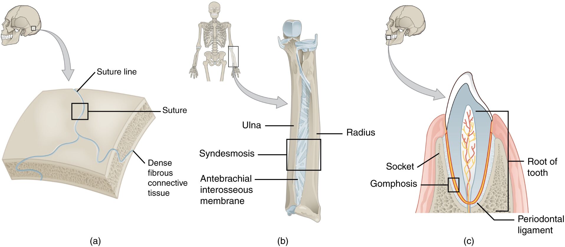
Suture Line
The suture line refers to the jagged, interlocking junction where cranial bones meet, forming an immovable joint in the skull. These lines, visible in the sutures, enhance stability by increasing the surface area of contact between bones.
Suture
The suture is a type of fibrous joint that connects the bones of the skull, such as the frontal and parietal bones, through dense fibrous connective tissue. It provides rigidity and protection to the brain by preventing movement between the cranial bones.
Dense Fibrous Connective Tissue
Dense fibrous connective tissue is the strong, collagen-rich material that binds the bones in fibrous joints like sutures, syndesmoses, and gomphoses. It ensures minimal movement, offering stability and strength to the joint structure.
Ulna
The ulna is one of the two long bones of the forearm, located on the side opposite the thumb, and is involved in a syndesmosis joint with the radius. It plays a key role in forearm stability and supports movements like pronation and supination.
Radius
The radius is the other long bone of the forearm, positioned on the same side as the thumb, and forms a syndesmosis with the ulna via the antebrachial interosseous membrane. It facilitates forearm rotation and weight transmission during hand movements.
Syndesmosis
The syndesmosis is a fibrous joint where bones, such as the radius and ulna, are connected by a sheet of dense fibrous tissue, specifically the antebrachial interosseous membrane. This joint allows slight movement while maintaining stability during forearm rotation.
Antepbrachial Interosseous Membrane
The antebrachial interosseous membrane is a broad sheet of dense fibrous connective tissue that connects the radius and ulna in a syndesmosis joint. It provides stability to the forearm and serves as an attachment site for muscles.
Gomphosis
The gomphosis is a specialized fibrous joint that anchors a tooth to its socket in the jawbone, using the periodontal ligament. This joint ensures the tooth remains securely in place while allowing slight movement to absorb chewing forces.
Socket
The socket, or alveolar socket, is the bony cavity in the jawbone that houses the root of a tooth in a gomphosis joint. It provides structural support and stability, ensuring the tooth can withstand the forces of biting and chewing.
Root of Tooth
The root of tooth is the portion of the tooth embedded in the jawbone’s socket, secured by the periodontal ligament in a gomphosis joint. It anchors the tooth firmly and contains nerves and blood vessels that nourish the tooth.
Periodontal Ligament
The periodontal ligament is a layer of dense fibrous connective tissue that connects the root of the tooth to the socket in a gomphosis joint. It acts as a shock absorber, distributing forces during chewing and maintaining tooth stability.
Anatomical Structure of Fibrous Joints
Types and Characteristics of Fibrous Joints
Fibrous joints are immovable or slightly movable joints connected by dense fibrous connective tissue, designed to provide stability in specific skeletal regions. They are categorized into sutures, syndesmoses, and gomphoses, each with unique structural features.
- The sutures of the skull, such as the coronal or sagittal sutures, interlock cranial bones with jagged edges, ensuring a rigid protective enclosure for the brain.
- The syndesmosis between the radius and ulna, formed by the antebrachial interosseous membrane, allows slight movement, supporting forearm rotation while maintaining stability.
- The gomphosis joint secures teeth to the jawbone using the periodontal ligament, allowing minimal movement to absorb chewing forces without compromising stability.
- Dense fibrous connective tissue in all fibrous joints is rich in collagen, providing tensile strength and resistance to separation.
- These joints lack a synovial cavity, distinguishing them from synovial joints and reinforcing their role in structural support rather than mobility.
Structural Components and Connections
The components of fibrous joints work together to ensure stability and strength, with each type adapted to its specific anatomical location. Understanding these components highlights their role in skeletal integrity.
- In sutures, the suture line’s interlocking pattern increases the surface area of contact, enhancing the joint’s resistance to mechanical stress.
- The antebrachial interosseous membrane in a syndesmosis joint acts as a flexible yet strong connection, allowing the radius and ulna to move slightly while remaining aligned.
- The periodontal ligament in a gomphosis joint contains collagen fibers that anchor the tooth’s root to the socket, distributing forces evenly during mastication.
- Dense fibrous connective tissue across all fibrous joints provides a durable, non-elastic connection that prevents excessive movement.
- The absence of cartilage or synovial fluid in fibrous joints ensures their rigidity, making them ideal for protective and stabilizing roles.
Physical Introduction to Fibrous Joints
Biomechanical Functions of Fibrous Joints
Fibrous joints are designed to prioritize stability over mobility, playing a critical role in protecting vital structures and maintaining skeletal alignment. Their biomechanical properties ensure they can withstand various forces without compromising function.
- The sutures in the skull absorb and distribute impact forces, protecting the brain from trauma during activities like running or accidental falls.
- The syndesmosis between the radius and ulna facilitates slight movement, allowing forearm rotation while preventing excessive separation of the bones.
- The gomphosis joint absorbs compressive forces during chewing, with the periodontal ligament distributing stress to prevent damage to the tooth or jawbone.
- Dense fibrous connective tissue in these joints provides high tensile strength, resisting stretching or tearing under mechanical stress.
- The immobility or limited mobility of fibrous joints ensures structural integrity in areas like the skull and jaw, where movement could be detrimental.
Role in Skeletal Stability and Support
Fibrous joints contribute significantly to the skeletal system by providing stable connections that support the body’s framework. Their role in maintaining alignment and protecting vital structures is essential for overall function.
- Sutures create a solid cranial vault, ensuring the brain remains protected within a rigid, unyielding structure.
- The syndesmosis joint between the radius and ulna supports forearm stability, allowing coordinated movements like writing or lifting objects.
- The gomphosis joint ensures teeth remain securely anchored in the jaw, enabling efficient mastication without dislodgement.
- These joints work in tandem with surrounding bones and muscles to maintain proper alignment and load distribution across the skeletal system.
- The fibrous nature of these joints allows them to act as anchor points for muscle attachments, enhancing their role in skeletal support.
Clinical Insights: Fibrous Joint Conditions
Common Disorders of Fibrous Joints
Fibrous joints, while inherently stable, can be affected by various conditions that impact their function or the structures they connect. Understanding these disorders is crucial for effective diagnosis and management.
- Craniosynostosis, a condition affecting sutures, involves the premature fusion of cranial sutures, leading to abnormal skull shapes and potential brain growth restrictions.
- Syndesmosis injuries, such as high ankle sprains, can occur in the forearm or ankle, where the interosseous membrane is stretched or torn, causing pain and instability.
- Periodontitis, a disease affecting the gomphosis joint, involves inflammation of the periodontal ligament and surrounding bone, potentially leading to tooth loss.
- Trauma to the skull can cause suture separation or diastasis, particularly in younger individuals where sutures are not fully ossified.
- Infections in the jaw, such as osteomyelitis, can weaken the gomphosis joint, compromising the stability of the tooth and surrounding bone.
Prevention and Management of Fibrous Joint Issues
Maintaining the health of fibrous joints is essential for preserving skeletal stability and function. Proactive measures and targeted interventions can help manage and prevent related issues.
- Early monitoring of cranial suture development in infants can detect craniosynostosis, allowing for surgical intervention to release fused sutures.
- Proper ankle and forearm support during sports can prevent syndesmosis injuries, such as using braces or taping to reduce stress on the interosseous membrane.
- Good oral hygiene practices, including regular brushing and flossing, can prevent periodontitis, protecting the periodontal ligament and gomphosis joint.
- Imaging studies, such as X-rays or MRIs, can diagnose suture separation or syndesmosis injuries, guiding appropriate treatment plans.
- In cases of severe periodontitis, dental interventions like scaling, root planing, or surgery may be necessary to preserve the gomphosis joint and tooth stability.
Conclusion
Fibrous joints, encompassing sutures, syndesmoses, and gomphoses, are vital for providing stability and support across the skeletal system, from the skull to the forearm and teeth. Their anatomical structure, characterized by dense fibrous connective tissue, ensures minimal movement while protecting critical structures like the brain and teeth. Understanding the roles of components like the suture line, antebrachial interosseous membrane, and periodontal ligament, along with potential conditions such as craniosynostosis or periodontitis, emphasizes the importance of maintaining their health. By prioritizing the care of fibrous joints, individuals can support overall skeletal integrity and function.

