This detailed diagram illustrates the critical early stages of human embryonic development, focusing on the formation and differentiation of the germ layers—ectoderm, mesoderm, and endoderm—following gastrulation. Understanding these fundamental processes is essential for comprehending how a single-celled zygote ultimately gives rise to the complex array of organs and systems that constitute a complete organism. This image provides a clear visual representation of the embryonic structures and their respective contributions to the developing fetus, highlighting the intricate orchestration of cellular migration and specialization during the third week of gestation and beyond.
Embryonic Structures and Their Roles:
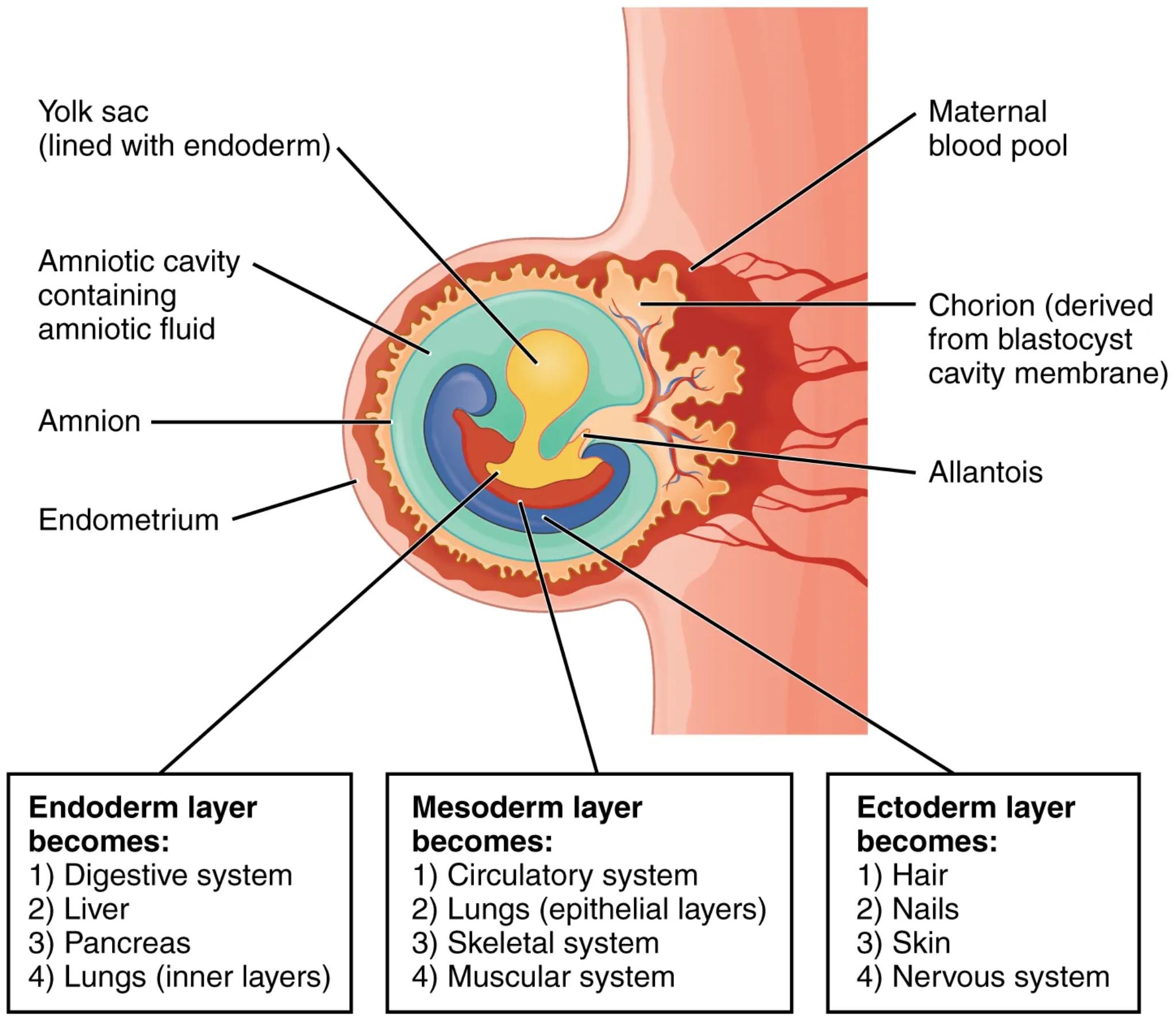
Yolk sac (lined with endoderm): The yolk sac is an extraembryonic membrane that plays a crucial role in early embryonic nutrition and blood cell formation. In humans, it is relatively small and primarily involved in the initial transfer of nutrients from the mother to the embryo before the placenta is fully functional, and it is also a site for the development of primordial germ cells.
Amniotic cavity containing amniotic fluid: This fluid-filled sac surrounds the developing embryo, providing a protective cushion against physical shock and trauma. The amniotic fluid also facilitates fetal movement, helps regulate temperature, and prevents the adherence of the amnion to the embryo.
Amnion: The amnion is the innermost extraembryonic membrane that encloses the embryo and the amniotic fluid. It is responsible for forming the amniotic sac, which creates the protective environment vital for proper fetal development.
Endometrium: The endometrium is the inner lining of the uterus, which thickens and becomes highly vascularized in preparation for implantation of the embryo. It provides the necessary nutritional support and attachment site for the developing blastocyst.
Maternal blood pool: This refers to the lacunae within the decidua basalis of the endometrium, which are filled with maternal blood. These pools facilitate the exchange of nutrients, oxygen, and waste products between the mother and the developing embryo via the chorionic villi.
Chorion (derived from blastocyst cavity membrane): The chorion is the outermost extraembryonic membrane that surrounds the embryo and other membranes. It develops into the fetal part of the placenta and is essential for nutrient and gas exchange between the mother and the fetus.
Allantois: The allantois is a diverticulum of the posterior wall of the yolk sac. In humans, it is a vestigial structure that contributes to the formation of the umbilical cord and the urinary bladder, and it is also involved in early blood formation.
Fates of the Germ Layers:
The differentiation of the three primary germ layers—ectoderm, mesoderm, and endoderm—is a pivotal event in embryogenesis, leading to the formation of all tissues and organs.
- Endoderm layer becomes:
- Digestive system: This includes the lining of the gastrointestinal tract, from the pharynx to the rectum, and associated glands.
- Liver: A vital organ involved in metabolism, detoxification, and bile production.
- Pancreas: An endocrine and exocrine gland producing hormones like insulin and digestive enzymes.
- Lungs (inner layers): The epithelial lining of the respiratory tract and the glandular tissue of the lungs originate from the endoderm.
- Mesoderm layer becomes:
- Circulatory system: This encompasses the heart, blood vessels, and blood cells, forming the body’s transport network.
- Lungs (epithelial layers): While the inner lining is endodermal, the connective tissue and muscle layers of the lungs are mesodermal in origin.
- Skeletal system: All bones, cartilage, and ligaments that provide structural support to the body are derived from the mesoderm.
- Muscular system: Both smooth and striated muscles throughout the body develop from the mesoderm, enabling movement and various physiological functions.
- Ectoderm layer becomes:
- Hair: Epidermal appendages that provide insulation and protection.
- Nails: Keratinized structures that protect the fingertips and toes.
- Skin: The epidermis, the outermost protective layer of the integumentary system, is ectodermal.
- Nervous system: This includes the brain, spinal cord, and peripheral nerves, which control and coordinate all bodily activities.
The journey from a fertilized egg to a fully formed organism is a testament to precise genetic programming and cellular communication. Following gastrulation, the three distinct germ layers are established, each possessing a unique developmental destiny. The ectoderm primarily gives rise to structures that interact with the external environment, such as the skin and nervous system, while the endoderm forms the linings of internal organs and glands. The mesoderm, positioned between the other two layers, differentiates into connective tissues, muscles, and the circulatory system, forming the bulk of the body’s structural and functional components. This intricate process of cell migration, differentiation, and organization underpins the development of every organ system, highlighting the remarkable complexity of early human embryology.
This comprehensive overview of embryonic germ layers and their ultimate fates provides a foundational understanding of developmental biology. By appreciating how these initial cellular divisions and differentiations lead to specialized tissues and complex organs, one can better grasp the interconnectedness of biological systems and the delicate balance required for healthy development. The diagram serves as an invaluable tool for visualizing these fundamental processes, offering insights into both typical development and the origins of congenital anomalies. The precision with which these layers unfold their developmental programs underscores the elegance and efficiency of biological design.

