The upper airway serves as the critical entry point for respiration, connecting the external environment to the lungs through a complex network of structures. This anatomical region, encompassing the nasal cavity, pharynx, and larynx, plays a pivotal role in filtering air, producing sound, and facilitating swallowing. A detailed examination of its components through sectional diagrams provides valuable insights into its functional design and clinical significance.
Key Anatomical Labels in the Diagram
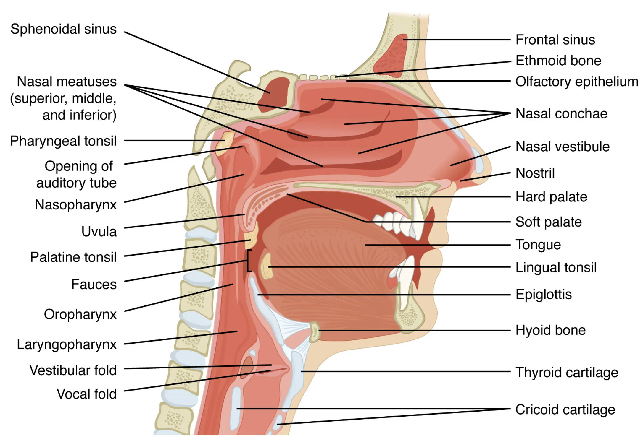
This section outlines each labeled component, offering a clear understanding of their roles and positions within the upper airway.
Sphenoidal sinus: The sphenoidal sinus is a paired air-filled cavity located within the sphenoid bone, situated behind the nasal cavity. It helps lighten the skull, resonates voice, and drains mucus into the nasal passages via the sphenoethmoidal recess.
Frontal sinus: Positioned within the frontal bone above the eyes, the frontal sinus contributes to skull weight reduction and humidifies inhaled air. It connects to the nasal cavity through the frontonasal duct, playing a role in sinus drainage.
Ethmoid bone: The ethmoid bone forms part of the nasal septum and lateral nasal walls, containing multiple air cells that filter and warm air. It also houses the olfactory receptors, essential for the sense of smell.
Olfactory epithelium: Located in the upper nasal cavity, the olfactory epithelium contains specialized cells that detect odor molecules, initiating the olfactory nerve signals. This thin layer is crucial for the perception of scents and flavors.
Nasal conchae: The nasal conchae, or turbinates, are scroll-like structures projecting from the lateral nasal walls, increasing surface area for air humidification and filtration. They also direct airflow to optimize respiratory efficiency.
Nasal vestibule: The nasal vestibule is the anterior entryway of the nasal cavity, lined with hair and mucus to trap dust and pathogens. It transitions into the nasal cavity proper, marking the initial stage of air filtration.
Nostril: The nostril, or external naris, serves as the external opening of the nasal cavity, allowing air intake and expulsion. It is supported by cartilage and skin, adapting to changes in airflow dynamics.
Hard palate: The hard palate forms the anterior roof of the mouth, separating the oral cavity from the nasal cavity with a bony structure. It provides a stable surface for chewing and supports speech articulation.
Soft palate: The soft palate is a muscular extension of the hard palate, elevating during swallowing to prevent food from entering the nasal cavity. It also plays a role in closing off the nasopharynx during phonation.
Tongue: The tongue occupies the oral cavity floor, aiding in food manipulation and taste perception through its papillae. It collaborates with the soft palate to guide food toward the pharynx during swallowing.
Lingual tonsil: Located at the tongue’s base, the lingual tonsil is part of the lymphatic ring protecting against infection. It can become inflamed, contributing to conditions like tonsillitis.
Epiglottis: The epiglottis is a cartilage flap at the larynx entrance, closing during swallowing to prevent food from entering the airway. It ensures a clear passage for air when open, coordinating with breathing.
Hyoid bone: The hyoid bone is a U-shaped structure below the mandible, supporting the tongue and larynx without direct skeletal articulation. It anchors muscles involved in swallowing and speech.
Thyroid cartilage: The thyroid cartilage forms the Adam’s apple, a prominent laryngeal structure protecting the vocal cords. It houses the vocal folds and supports thyroid hormone production by the thyroid gland, releasing T3 and T4.
Cricoid cartilage: The cricoid cartilage is a ring-like structure below the thyroid cartilage, forming the larynx’s base. It provides structural support and connects to the trachea, ensuring airway stability.
Pharyngeal tonsil: The pharyngeal tonsil, or adenoid, is located near the nasopharynx opening, contributing to immune defense by trapping pathogens. Its enlargement can obstruct breathing, particularly in children.
Opening of auditory tube: The opening of the auditory tube, or Eustachian tube, connects the middle ear to the nasopharynx, equalizing ear pressure. It also drains mucus, preventing ear infections.
Nasopharynx: The nasopharynx is the upper pharyngeal region behind the nasal cavity, serving as an airway passage. It houses the pharyngeal tonsil and auditory tube openings, facilitating breathing and pressure regulation.
Nasal meatuses (superior, middle, and inferior): The nasal meatuses are air passages beneath the nasal conchae, channeling airflow for filtration and humidification. They connect to the paranasal sinuses, aiding in mucus drainage.
Uvula: The uvula is a small projection at the soft palate’s posterior end, assisting in closing the nasopharynx during swallowing. It also contributes to speech by altering airflow.
Palatine tonsil: The palatine tonsil lies on each side of the oropharynx, forming part of the lymphatic ring against infection. Inflammation here can lead to sore throats or tonsillectomy considerations.
Fauces: The fauces is the passageway between the oral cavity and oropharynx, marked by the palatine arches. It directs food and air, coordinating swallowing and breathing.
Oropharynx: The oropharynx extends from the soft palate to the epiglottis, serving as a common pathway for air and food. It contains the palatine and lingual tonsils, supporting immune function.
Laryngopharynx: The laryngopharynx, or hypopharynx, lies below the oropharynx, transitioning to the esophagus and larynx. It directs food to the stomach and air to the trachea during respiration.
Vestibular fold: The vestibular fold, or false vocal cord, lies above the true vocal cord, protecting the airway. It plays a minor role in phonation but aids in closing the larynx during swallowing.
Vocal fold: The vocal fold, or true vocal cord, produces sound by vibrating as air passes, modulated by muscle tension. It is innervated by the recurrent laryngeal nerve, critical for voice control.
The Bony and Cartilaginous Framework
The upper airway’s skeletal elements provide a robust yet flexible structure. This foundation supports respiratory and phonatory functions effectively.
- The sphenoidal and frontal sinuses reduce skull weight, resonating sound during speech.
- The ethmoid bone’s air cells enhance olfaction and sinus drainage.
- The hyoid bone anchors the larynx, facilitating tongue movement.
- Thyroid and cricoid cartilages protect the vocal apparatus, ensuring clear communication.
- These structures adapt to pressure changes, maintaining airway patency.
Soft Tissue Contributions to Airway Function
Soft tissues enhance the upper airway’s versatility, adapting to diverse physiological demands. Their coordination is key to seamless operation.
- The soft palate elevates to seal the nasopharynx, preventing nasal reflux.
- Nasal conchae increase air turbulence, trapping particles effectively.
- The epiglottis and vestibular folds safeguard the larynx during swallowing.
- The tongue and uvula guide food, supporting digestive initiation.
- Mucous membranes humidify air, protecting delicate tissues from drying.
Physiological Roles in Respiration and Phonation
The upper airway’s design optimizes breathing and sound production. Its anatomy supports a range of vital processes.
- Nasal vestibule and meatuses filter air, removing debris before lung entry.
- The larynx, with vocal folds, generates pitch and volume for speech.
- The pharyngeal regions coordinate swallowing and breathing transitions.
- Olfactory epithelium detects scents, linking to the brain’s limbic system.
- Pressure equalization via the auditory tube prevents ear discomfort.
Clinical Considerations and Anatomical Variations
Understanding airway anatomy aids in diagnosing and treating related conditions. Variations can influence treatment approaches.
- Sinus infections often involve the sphenoidal and frontal sinuses, requiring drainage.
- Tonsil hypertrophy, including pharyngeal and palatine types, may obstruct breathing.
- Vocal fold paralysis affects phonation, linked to recurrent laryngeal nerve damage.
- Laryngopharyngeal reflux can irritate the hypopharynx, causing chronic cough.
- Imaging like CT scans reveals anatomical deviations for surgical planning.
The upper airway’s intricate design reflects its multifaceted role in respiration, phonation, and immune defense. By studying its components through detailed sectional views, one gains a deeper understanding of its contributions to human health, highlighting the elegance of this vital system.

