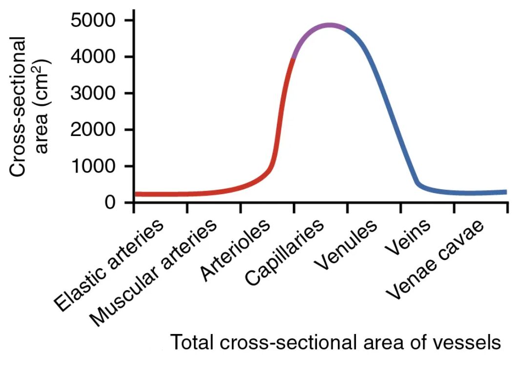
The total cross-sectional area of vessels is a critical factor in understanding how blood flows through the circulatory system, influencing velocity, pressure, and exchange efficiency. This diagram illustrates the progressive changes in cross-sectional area from large arteries to tiny capillaries and back to veins, highlighting the anatomical and physiological implications for vascular function.
Aorta Aorta has a total cross-sectional area of approximately 4-5 cm², reflecting its role as the primary outlet for blood from the heart. Its limited area supports high-pressure, rapid flow to distribute blood to the systemic circulation.
Elastic arteries Elastic arteries, such as the carotid and subclavian, collectively have a cross-sectional area of about 20 cm² due to their branching nature. This increase aids in smoothing out pulsatile flow and maintaining steady pressure downstream.
Muscular arteries Muscular arteries, including the brachial and femoral, contribute a total cross-sectional area of around 40 cm² as they further divide. This area supports controlled distribution to specific organs, adjusting flow based on demand.
Arterioles Arterioles have a combined cross-sectional area of approximately 50-60 cm², reflecting their numerous branches. This expansion begins to slow blood velocity, preparing it for capillary exchange.
Capillaries Capillaries boast the largest total cross-sectional area, estimated at 4,500-5,000 cm² due to their vast numbers. This dramatic increase reduces flow speed, optimizing the time for nutrient and gas exchange with tissues.
Venules Venules have a total cross-sectional area of about 200-300 cm² as they collect blood from capillaries. This reduction in area compared to capillaries facilitates the transition to larger venous vessels.
Veins Veins possess a total cross-sectional area of approximately 500 cm², reflecting their role as capacitance vessels. This large area allows them to store a significant portion of the body’s blood volume.
Venae cavae Venae cavae have a total cross-sectional area of about 10-15 cm², narrowing as they deliver blood to the heart. This reduction supports the low-pressure return of deoxygenated blood to the right atrium.
Overview of Total Cross-Sectional Area
This diagram showcases how the total cross-sectional area of vessels varies across the circulatory system. These changes are essential for regulating blood flow and supporting physiological needs.
- Aorta starts with a small area, driving high-velocity flow.
- Elastic arteries and muscular arteries increase area gradually, distributing blood.
- Arterioles mark a transition with a moderate area increase.
- Capillaries reach a peak area, enhancing exchange efficiency.
- Venules, veins, and venae cavae reduce area, aiding venous return.
Anatomical Role of Cross-Sectional Area
The total cross-sectional area of vessels reflects their specialized functions within the circulatory network. This variation ensures effective blood distribution and exchange.
- Aorta area supports the initial high-pressure ejection from the left ventricle.
- Elastic arteries area cushions the pulse, reducing stress on smaller vessels.
- Muscular arteries area allows selective flow to organs like the brain or liver.
- Arterioles area slows flow, preparing blood for capillary beds.
- Capillaries vast area maximizes contact with tissues for oxygen delivery.
Microcirculation and Capillary Area
Capillaries dominate with their extensive cross-sectional area in the microcirculation. This feature is critical for the exchange process.
- Capillaries area supports a network of 10-20 billion vessels in the body.
- The large area reduces velocity to 0.3 cm/sec, enhancing diffusion time.
- Arterioles area feeds into this network, controlling entry volume.
- Precapillary sphincters adjust capillaries area based on metabolic demand.
- This setup ensures efficient nutrient uptake and waste removal.
Venous Return and Larger Vessel Area
Veins and venae cavae utilize their cross-sectional area for low-pressure return. This design supports the body’s circulatory balance.
- Venules area consolidates flow from capillaries, reducing turbulence.
- Veins area holds 60-70% of blood volume, acting as reservoirs.
- Venae cavae area narrows to direct blood into the heart efficiently.
- Valves within veins prevent backflow despite the large area.
- Muscle pump action leverages this area to enhance venous return.
Physical Implications of Cross-Sectional Area
The total cross-sectional area influences flow velocity and resistance according to Poiseuille’s law. These dynamics are vital for circulatory health.
- Aorta small area drives a velocity of 35 cm/sec under high pressure.
- Elastic arteries area slows velocity to 20-30 cm/sec, stabilizing flow.
- Muscular arteries area maintains velocity while directing blood.
- Arterioles area reduces velocity to 14-21 cm/sec, increasing resistance.
- Capillaries large area drops velocity significantly, aiding exchange.
Clinical Relevance of Cross-Sectional Area
Changes in the total cross-sectional area of vessels can indicate health issues. Monitoring these variations guides clinical interventions.
- Reduced capillaries area may impair perfusion in conditions like diabetes.
- Enlarged veins area can lead to varicose veins due to valve failure.
- Narrowed arterioles area is associated with hypertension or atherosclerosis.
- Increased aorta area might suggest an aneurysm, requiring imaging.
- Adjusting muscular arteries area with vasodilators treats circulatory deficits.
In conclusion, the total cross-sectional area of vessels diagram illustrates the critical roles of aorta, elastic arteries, muscular arteries, arterioles, capillaries, venules, veins, and venae cavae in regulating blood flow and exchange. This understanding of how area impacts velocity and pressure deepens insight into circulatory efficiency. It also provides a foundation for addressing vascular conditions with targeted care.

