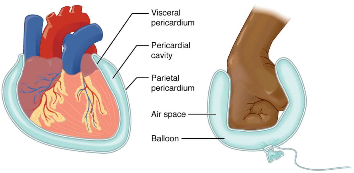
The human body relies on specialized membranes to protect and support its vital organs, with the serous membrane playing a critical role in this process. This image illustrates the Visceral Pericardium, Pericardial Cavity, Parietal Pericardium, Air Space, and Balloon, using a heart model and a balloon analogy to demonstrate how the serous membrane surrounds the heart. Understanding this structure enhances knowledge of cardiac anatomy and its protective mechanisms.
Label Introductions:
- Visceral Pericardium: The visceral pericardium is the inner layer of the serous membrane, directly covering the heart’s surface. It provides a smooth, protective coating that reduces friction during heartbeats.
- Pericardial Cavity: The pericardial cavity is a small space between the visceral and parietal pericardium, filled with pericardial fluid. This fluid lubricates the heart, allowing it to move freely within the cavity.
- Parietal Pericardium: The parietal pericardium forms the outer layer of the serous membrane, lining the pericardial sac. It anchors the heart to surrounding structures while offering additional protection.
- Air Space: The air space in the balloon analogy represents the pericardial cavity, illustrating the fluid-filled gap. It highlights how the membrane layers create a cushioning effect around the organ.
- Balloon: The balloon serves as a model, mimicking the double-layered structure of the serous membrane around a fist, akin to the heart. It demonstrates how the membrane folds to envelop and protect the organ.
Overview of Serous Membrane Anatomy
The serous membrane is a thin, double-layered structure that lines body cavities and covers organs, ensuring smooth movement and protection. The pericardial cavity, lined by the visceral and parietal pericardium, is a prime example, safeguarding the heart from friction and trauma. This membrane system is essential for maintaining organ function across various body systems.
- Reduces friction between organs and cavity walls during movement.
- Produces serous fluid to lubricate and cushion vital structures.
- Protects organs from infection and mechanical damage.
- Facilitates the study of organ relationships in medical imaging.
The Pericardium: Heart’s Protective Layers
The pericardium consists of the visceral pericardium and parietal pericardium, forming a protective sac around the heart. The visceral layer adheres closely to the heart muscle, while the parietal layer forms the outer boundary, creating the pericardial cavity. This dual-layer design is crucial for cardiac efficiency.
- The visceral pericardium contains blood vessels that nourish the heart.
- The pericardial cavity holds about 15-50 mL of fluid for lubrication.
- The parietal pericardium is attached to the diaphragm and sternum.
- Inflammation here, known as pericarditis, can affect heart function.
Pericardial Cavity: Fluid Dynamics
The pericardial cavity is a narrow space filled with serous fluid, acting as a lubricant between the pericardial layers. This fluid prevents adhesion during the heart’s constant motion, mimicking the air space in the balloon analogy. It ensures the heart operates without excessive resistance.
- Maintains a low-pressure environment around the heart.
- Excess fluid accumulation can lead to pericardial effusion.
- The fluid is produced by both pericardial layers.
- Supports the heart’s rhythmic contractions and relaxations.
Balloon Analogy: Visualizing the Structure
The balloon analogy effectively illustrates the serous membrane’s function, with the fist representing the heart. The balloon’s inner and outer layers correspond to the visceral and parietal pericardium, with the air space symbolizing the pericardial cavity. This model simplifies the concept of membrane folding and protection.
- The balloon’s elasticity mimics the pericardium’s adaptability.
- The air space shows how fluid prevents direct contact.
- The fist’s movement parallels the heart’s beating action.
- This analogy aids in teaching complex anatomical relationships.
Clinical Significance of the Serous Membrane
The serous membrane’s structure, particularly around the heart, has significant clinical implications. Conditions like pericarditis can cause inflammation of the pericardium, leading to chest pain and fluid buildup. Understanding the visceral pericardium and parietal pericardium helps in diagnosing and treating such disorders.
- Pericarditis may result from infections or autoimmune diseases.
- Fluid in the pericardial cavity can compress the heart, causing tamponade.
- Imaging techniques like echocardiography assess membrane health.
- Surgical interventions may address severe pericardial issues.
Conclusion
The serous membrane, exemplified by the pericardial cavity and its layers, is a remarkable feature of human anatomy that protects the heart. The visceral pericardium, parietal pericardium, and their fluid-filled space work together to ensure smooth cardiac function, much like the balloon analogy suggests. This knowledge not only deepens anatomical understanding but also supports effective management of related medical conditions.

