The semicircular canals, a vital part of the inner ear’s vestibular system, are key to sensing rotational movements of the head, ensuring balance and spatial awareness. This intricate mechanism involves the cupula and hair cells, which respond to fluid shifts within the canals, providing critical data for coordinating head and eye movements.
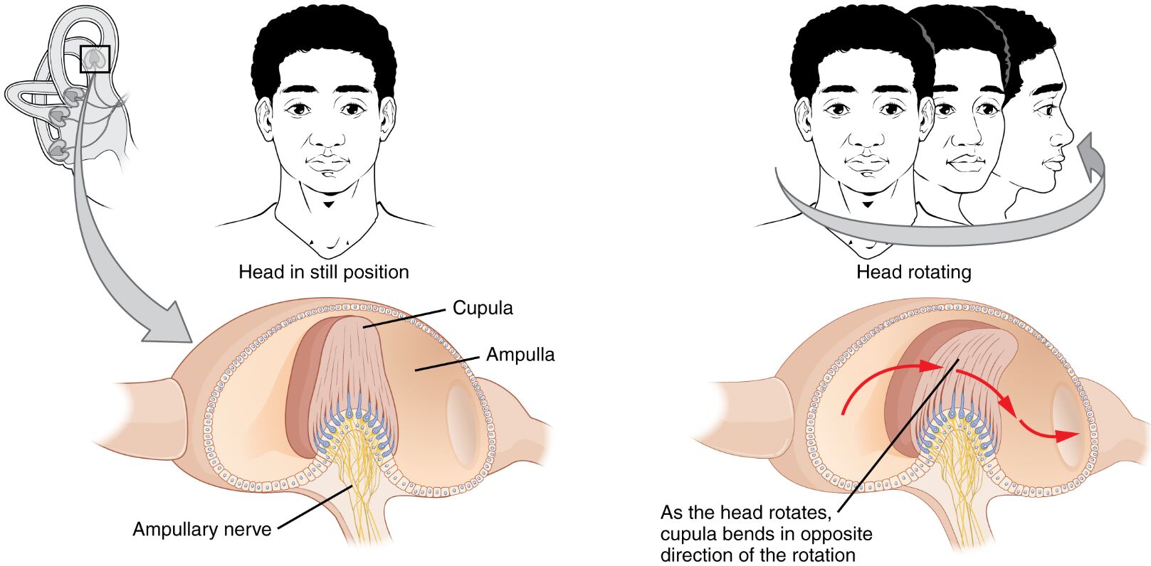
Cupula The cupula is a gelatinous structure within the ampulla of the semicircular canals, housing hair cell stereocilia. It bends in response to endolymph movement during head rotation, triggering neural signals that indicate the direction of movement.
Ampulla The ampulla is the widened section at the base of each semicircular canal, containing the cupula and hair cells. It serves as the sensory hub where rotational acceleration is detected and converted into electrical impulses.
Ampullary nerve The ampullary nerve is a branch of the vestibular division of the vestibulocochlear nerve, transmitting signals from the hair cells in the ampulla. It relays information about head rotation to the brainstem for processing and balance adjustment.
Anatomy of the Semicircular Canals
The semicircular canals are three fluid-filled loops arranged orthogonally in the inner ear, designed to detect angular acceleration. Their unique structure allows for precise detection of head rotation in all planes.
- Each canal is oriented at approximately 90 degrees to the others, forming the superior, posterior, and lateral canals.
- The canals are filled with endolymph, a fluid that moves in response to head motion, interacting with the cupula.
- The ampulla at one end of each canal houses the sensory epithelium, rich in hair cells and supporting cells.
- Membranous ducts connect the canals to the utricle, facilitating fluid communication within the vestibular system.
- The bony labyrinth encasing the canals provides structural support and protection.
- Blood vessels and nerves, including the ampullary nerve, nourish and innervate this region.
Physiology of Rotational Detection
Rotational movement triggers a dynamic response within the semicircular canals, where fluid inertia plays a central role. As the head rotates, the endolymph lags, causing the cupula to deflect and activate hair cells.
- The cupula acts as a pivot point, bending opposite to the direction of head rotation due to endolymph flow.
- Hair cells within the ampulla depolarize or hyperpolarize based on the direction of cupula deflection.
- This deflection opens ion channels, allowing potassium influx and generating action potentials in afferent nerves.
- The three pairs of canals (left and right for each plane) work in opposition, enhancing directional accuracy.
- Adaptation occurs over time as the endolymph stabilizes, reducing sensitivity to sustained rotation.
- The vestibular system integrates these signals with visual input for coordinated responses.
Role of the Cupula in Sensing Motion
The cupula serves as the mechanical link between fluid movement and sensory transduction in the semicircular canals. Its elasticity and position make it ideal for detecting angular changes.
- Composed of a glycoprotein matrix, the cupula is semi-rigid yet flexible, allowing controlled bending.
- During rotation, endolymph pushes against the cupula, displacing it by up to 10 degrees depending on acceleration.
- Hair cell stereocilia embedded in the cupula respond to this displacement, with taller cilia near the kinocilium for polarity.
- The cupula resets to its neutral position after rotation stops, aided by elastic recoil and fluid dynamics.
- Damage to the cupula, such as in trauma, can lead to vertigo, though this diagram shows normal function.
- Its sensitivity peaks at frequencies between 0.1 and 5 Hz, aligning with natural head movements.
Hair Cells and Neural Transmission
Hair cells within the ampulla are the sensory transducers, converting mechanical stimuli into neural signals. Their stereocilia bundles are finely tuned to detect cupula movement.
- Stereocilia are linked by tip links, which open potassium channels upon bending toward the kinocilium.
- Type I hair cells, enveloped by calyx terminals, provide rapid, high-fidelity signals for dynamic motion.
- Type II hair cells, with bouton endings, contribute to sustained responses and modulation.
- The ampullary nerve carries these signals via myelinated fibers to the vestibular nuclei.
- Efferent fibers from the brainstem regulate hair cell sensitivity, preventing overstimulation.
- This process supports reflexes like the vestibulo-ocular reflex, stabilizing gaze during head turns.
Integration with the Vestibular System
Signals from the semicircular canals are processed centrally to maintain equilibrium and coordinate movements. The ampullary nerve connects this peripheral system to higher brain centers.
- The vestibular nuclei in the brainstem receive input from all six canals, integrating bilateral data.
- Projections to the cerebellum refine motor adjustments, such as during rapid head turns.
- The medial longitudinal fasciculus links to ocular motor nuclei for eye movement synchronization.
- Vestibulospinal tracts influence neck and limb muscles for postural stability.
- Cortical areas process conscious perception of rotation, aiding spatial navigation.
- Dysfunction in this pathway can cause nystagmus, but the diagram illustrates healthy anatomy.
Clinical Insights into Rotational Sensing
Understanding the semicircular canals aids in diagnosing balance disorders, though this image focuses on normal physiology. Conditions like vestibular neuritis can affect ampullary nerve function.
- Caloric testing uses temperature changes to stimulate the canals, assessing their response.
- Vestibular rehabilitation exercises enhance canal adaptation in cases of imbalance.
- Studies on astronauts explore canal function in microgravity, where rotational cues diminish.
- Pharmacotherapy may target hair cell activity to alleviate vertigo symptoms.
- Imaging techniques like CT scans visualize canal anatomy for diagnostic purposes.
In conclusion, the semicircular canals exemplify the body’s remarkable ability to detect and respond to rotational movements. Through the interplay of the cupula, hair cells, and ampullary nerve, this system ensures precise balance and orientation, underpinning our interaction with the environment.

