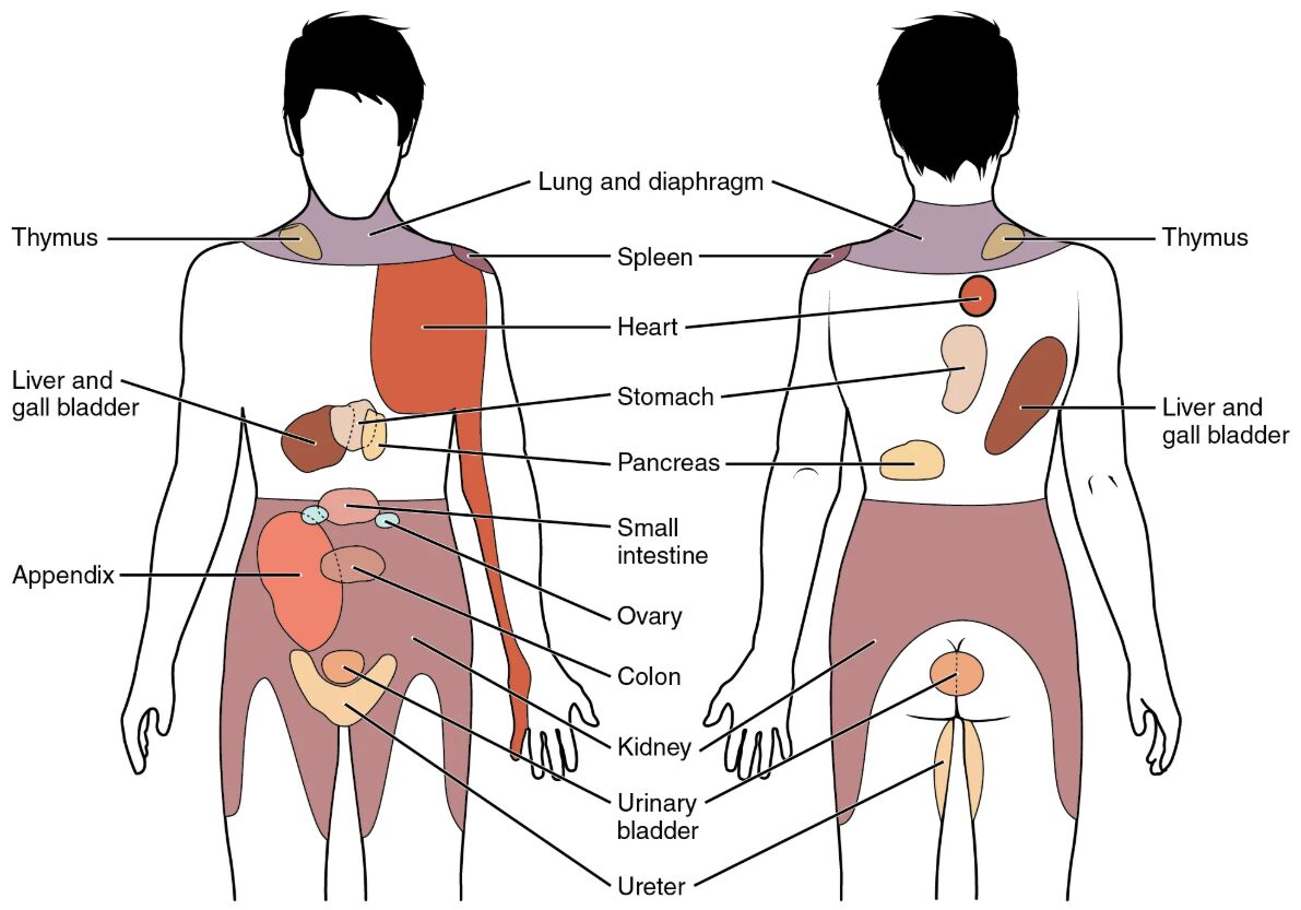
The referred pain chart provides a fascinating glimpse into how the body interprets sensations from internal organs, mapping them onto specific surface areas despite their distant origins. This diagram illustrates the complex neural connections that lead to the phenomenon of referred pain, where discomfort from organs like the heart or kidneys is perceived in regions such as the shoulder or lower back. Understanding these patterns enhances clinical diagnosis and treatment, offering valuable insights into the interplay between visceral and somatic nervous systems.
Labeled Components in the Diagram
Thymus The thymus is depicted in the upper chest and neck region, playing a key role in immune system development by producing T-lymphocytes. Referred pain from this area may be felt in the upper chest, often linked to inflammation or pressure on adjacent structures.
Liver and gall bladder The liver and gall bladder, located in the upper right abdomen, are involved in digestion and bile production. Pain from these organs can refer to the right shoulder or upper back due to shared nerve pathways with the diaphragm.
Lung and diaphragm The lung and diaphragm, shown in the chest area, facilitate breathing and separate the thoracic and abdominal cavities. Referred pain here might manifest in the shoulder or neck, especially in cases of pleurisy or diaphragmatic irritation.
Spleen The spleen, positioned in the upper left abdomen, filters blood and supports immune function. Discomfort from the spleen can refer to the left shoulder, a phenomenon often observed in splenic rupture or enlargement.
Heart The heart, centrally located in the chest, pumps blood throughout the body and relies on coronary circulation. Referred pain from cardiac issues, such as angina, commonly radiates to the left arm or jaw.
Stomach The stomach, found in the upper left abdomen, aids in digestion through acid and enzyme secretion. Pain from gastric ulcers or gastritis may refer to the upper abdominal or chest region.
Pancreas The pancreas, situated behind the stomach, regulates blood sugar and aids digestion with enzymes. Referred pain from pancreatitis often extends to the back or upper abdomen.
Small intestine The small intestine, located in the central lower abdomen, absorbs nutrients from digested food. Discomfort here might refer to the mid-abdominal area due to its extensive nerve supply.
Ovary The ovary, positioned in the pelvic region, produces eggs and hormones like estrogen. Ovarian pain can refer to the lower abdomen or back, especially during conditions like ovarian cysts.
Colon The colon, extending through the abdomen and pelvis, reabsorbs water and forms feces. Referred pain from colitis or diverticulitis may be felt in the lower abdomen or flanks.
Appendix The appendix, located in the lower right abdomen, is a small pouch with an unclear primary function. Appendicitis often causes referred pain to the navel or right lower quadrant.
Kidney The kidney, situated in the mid-back on either side, filters blood to produce urine. Kidney stones or infections can refer pain to the lower back or groin.
Urinary bladder The urinary bladder, located in the pelvic cavity, stores urine before excretion. Bladder irritation or infection may cause referred pain in the lower abdomen or perineum.
Ureter The ureter, running from the kidney to the bladder, transports urine. Obstructions like kidney stones can lead to referred pain along its path to the groin.
Anatomy of Referred Pain
Referred pain arises due to the convergence of visceral and somatic afferent fibers in the spinal cord. This overlap confuses the brain’s perception, attributing visceral pain to somatic regions.
- The phenomenon relies on shared dermatomes, which are areas of skin innervated by the same spinal nerves as internal organs.
- Visceral pain signals travel via sympathetic nerves, often activating the same second-order neurons as somatic input.
- The thoracic and lumbar spinal segments are particularly prone to this convergence, explaining common referral patterns.
- Conditions like myocardial infarction exemplify how heart pain radiates to the left arm due to T1-T4 dermatomes.
- This mapping aids clinicians in diagnosing internal issues based on external pain locations.
Physiological Basis of Referred Pain
The physiological mechanism involves the central nervous system’s interpretation of sensory input from visceral organs. This process highlights the complexity of neural integration and pain perception.
- Visceral afferents enter the spinal cord through the dorsal root ganglia, often alongside somatic fibers.
- The brain lacks a direct map of visceral structures, leading to misattribution of pain signals.
- Inflammatory mediators, such as prostaglandins, amplify visceral nociceptor activity, enhancing referral.
- Sympathetic activation during stress or injury can intensify perceived pain in referred areas.
- Experimental studies using capsaicin injections demonstrate how visceral stimulation triggers somatic discomfort.
Clinical Applications in Diagnosis
Understanding referred pain patterns is crucial for accurate medical diagnosis and effective treatment planning. This knowledge bridges anatomical insights with practical healthcare applications.
- Cardiac pain referral to the jaw or arm prompts immediate evaluation for heart conditions.
- Right shoulder pain may indicate gallbladder issues, guiding imaging like ultrasound.
- Lower back pain from kidney stones necessitates urinalysis and CT scans.
- Ovarian pain referral can lead to gynecological exams for cysts or torsion.
- Appendicitis diagnosis often begins with assessing navel-to-right-lower-quadrant pain migration.
Common Conditions Associated with Referred Pain
Various organ dysfunctions can trigger referred pain, each with distinct clinical presentations. Recognizing these patterns enhances patient care and outcomes.
- Gallstones cause biliary colic, with pain radiating to the right shoulder due to diaphragmatic irritation.
- Pancreatitis presents with epigastric pain referring to the back, often with nausea.
- Renal colic from ureteral obstruction radiates from the flank to the groin.
- Pleuritic chest pain may refer to the shoulder in lung infections or pneumothorax.
- Gastric reflux can mimic heart pain, requiring differential diagnosis with ECG.
Educational Value of the Referred Pain Chart
The referred pain chart serves as an invaluable tool for learning the body’s sensory mapping. It connects theoretical anatomy with practical clinical observations.
- Medical professionals use this chart to teach the relationship between internal organs and surface pain.
- It highlights the importance of thorough patient history in pinpointing pain origins.
- Workshops often include case studies to practice identifying referral patterns.
- The diagram supports understanding of neural plasticity in chronic pain conditions.
- Interactive models based on this chart enhance hands-on learning experiences.
Advances in Pain Management
Recent research explores targeted therapies to address referred pain, improving patient comfort and recovery. Innovations in this field offer hope for better outcomes.
- Nerve blocks target spinal segments to interrupt pain referral pathways.
- Pharmacological agents like gabapentin modulate central sensitization.
- Biofeedback techniques help patients manage perceived pain intensity.
- Surgical interventions, such as sympathectomy, address severe visceral pain cases.
- Ongoing studies investigate neuromodulation for chronic referral syndromes.
The referred pain chart underscores the intricate relationship between our internal organs and external sensations, offering a window into the body’s communication system. By mastering these patterns, healthcare providers can enhance diagnostic accuracy and tailor treatments to individual needs, ultimately improving patient well-being through a deeper understanding of human physiology.

