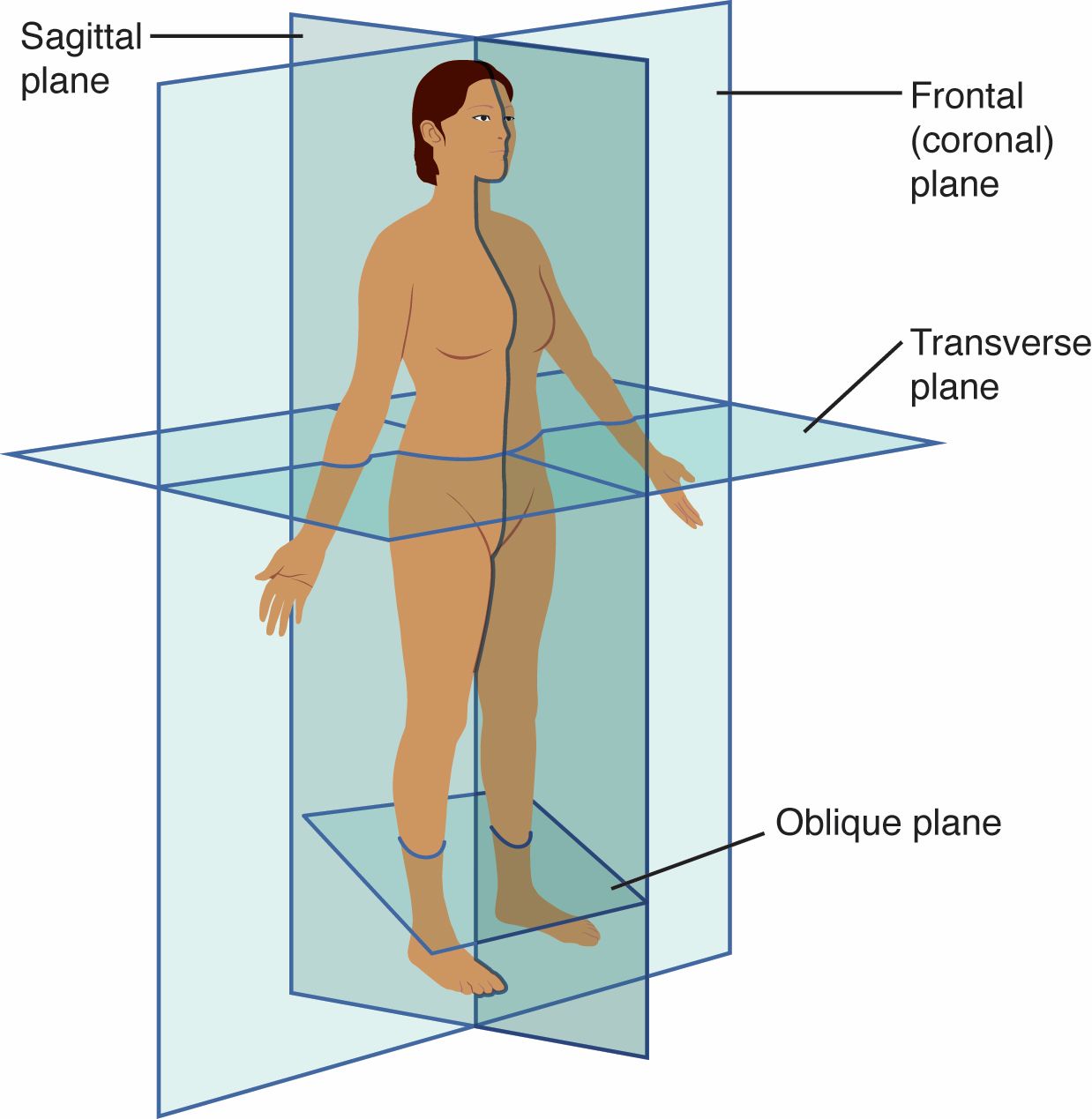
The human body is a marvel of structure and function, often analyzed through specific planes to understand its anatomy and aid in medical imaging. This image illustrates the Sagittal Plane, Frontal (Coronal) Plane, Transverse Plane, and Oblique Plane, which are essential for visualizing the body’s orientation and sections. Delving into these planes offers valuable insights into how professionals study and treat the human form with precision.
Label Introductions:
- Sagittal Plane: The sagittal plane divides the body into left and right sections, running vertically from front to back. It is commonly used to examine the lateral aspects of organs and structures, such as the brain or spine.
- Frontal (Coronal) Plane: The frontal, or coronal, plane separates the body into anterior (front) and posterior (back) portions, extending vertically. This plane is vital for imaging the chest, abdomen, and head, revealing relationships between front and back structures.
- Transverse Plane: The transverse plane cuts the body into superior (upper) and inferior (lower) parts, running horizontally across the body. It is frequently utilized in CT scans and surgeries to assess cross-sectional anatomy, such as the torso or limbs.
- Oblique Plane: The oblique plane runs at an angle, intersecting the body diagonally rather than straight. This plane is less common but useful for studying structures at unconventional angles, providing a unique perspective in complex imaging.
Understanding Anatomical Planes
Anatomical planes serve as foundational tools in medical science, guiding the study and visualization of the body’s internal layout. These planes allow for a systematic approach to dissecting and imaging, ensuring accurate diagnosis and treatment. Mastery of these planes enhances the ability to interpret medical scans and plan surgical interventions effectively.
- Provide a three-dimensional framework for analyzing body structures.
- Enable precise communication among healthcare professionals using standardized terms.
- Facilitate the creation of detailed anatomical models and educational resources.
- Support advanced imaging techniques like MRI and X-rays for diagnostic purposes.
Sagittal Plane: Dividing Left and Right
The Sagittal Plane offers a side view of the body, splitting it into symmetrical or asymmetrical halves. This plane is particularly useful for studying the midline structures, such as the nasal cavity or vertebral column. It helps in assessing conditions that affect one side of the body more than the other.
- Commonly used in neurosurgery to evaluate brain hemispheres.
- Allows visualization of the spinal cord’s alignment and potential deformities.
- Aids in studying the digestive tract’s lateral positioning.
- Essential for planning treatments involving unilateral injuries.
Frontal (Coronal) Plane: Front and Back Perspectives
The Frontal (Coronal) Plane provides a front-to-back view, ideal for examining the body’s vertical alignment. This plane is critical for imaging the lungs, heart, and abdominal organs, offering insights into their anterior-posterior relationships. It supports the diagnosis of conditions affecting the chest or pelvic regions.
- Key in assessing the heart’s position relative to the lungs.
- Used to evaluate the alignment of the spine in scoliosis cases.
- Helps in planning thoracic surgeries with precision.
- Reveals the layering of muscles and tissues in the abdominal wall.
Transverse Plane: Horizontal Cross-Sections
The Transverse Plane creates a top-down or bottom-up view, slicing the body horizontally. This plane is indispensable in cross-sectional imaging, such as identifying the liver’s location or assessing limb fractures. It provides a clear picture of organ distribution across different levels of the body.
- Critical for interpreting CT scans of the abdomen and pelvis.
- Used to measure the diameter of blood vessels in the torso.
- Assists in locating tumors or lesions in horizontal layers.
- Supports orthopedic evaluations of joint cross-sections.
Oblique Plane: Angled Insights
The Oblique Plane adds versatility by cutting the body at an angle, capturing details that other planes might miss. This plane is valuable in specialized imaging, such as evaluating the kidneys or joints from a diagonal perspective. It enhances the understanding of complex anatomical relationships.
- Useful in imaging the kidneys’ position relative to the spine.
- Applied in dental imaging to assess jaw angles.
- Aids in studying the pelvis from unconventional viewpoints.
- Supports detailed analysis in trauma cases with irregular injuries.
Conclusion
The exploration of anatomical planes like the Sagittal Plane, Frontal (Coronal) Plane, Transverse Plane, and Oblique Plane is crucial for a thorough understanding of human anatomy. These planes not only guide medical imaging but also underpin surgical planning and educational studies. By mastering their application, professionals can enhance diagnostic accuracy and improve patient outcomes, making them indispensable tools in the field of medicine.

