The olfactory system, a key player in the human sense of smell, relies on the intricate structure of the olfactory epithelium to detect and process odors from the environment. This image highlights the critical role of olfactory receptor neurons within this specialized tissue, offering a window into the initial stages of olfactory perception. This article provides a comprehensive examination of the anatomical and physiological aspects of this system, shedding light on its importance in sensory processing.
Labeled Parts of the Olfactory System
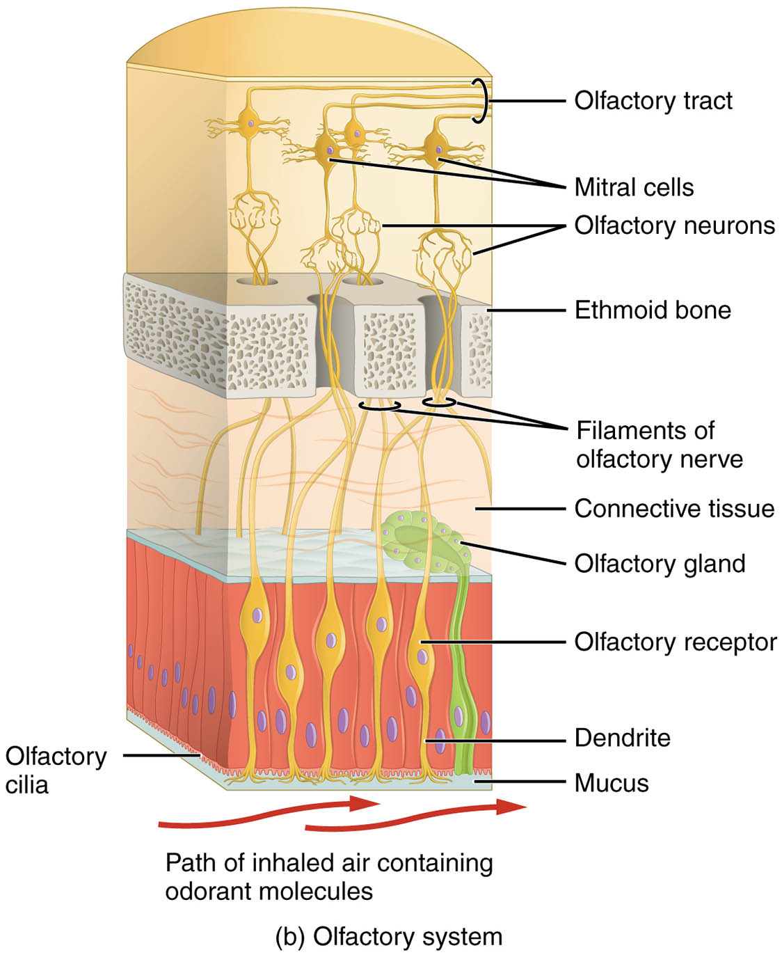
Olfactory epithelium The olfactory epithelium is a thin, specialized tissue lining the nasal cavity that contains olfactory receptor neurons responsible for detecting odors. It is supported by a layer of mucus and connective tissue, which helps trap and present odorant molecules to the receptors.
Olfactory receptor neuron The olfactory receptor neuron is a bipolar sensory cell within the olfactory epithelium, featuring dendrites with cilia that detect odorant molecules. Its axon extends to the olfactory bulb, transmitting sensory signals as part of the first cranial nerve.
Cilia Cilia are hair-like structures extending from the dendrites of olfactory receptor neurons, increasing the surface area for odorant detection. They move within the mucus layer, enhancing the interaction between odorants and receptors to initiate smell perception.
Mucus layer The mucus layer covers the olfactory epithelium, providing a moist environment that dissolves odorant molecules for detection. It is secreted by underlying glands, protecting the epithelium while facilitating the olfactory process.
Supporting cell The supporting cell, or sustentacular cell, is a non-neuronal cell within the olfactory epithelium that provides structural and metabolic support to olfactory receptor neurons. It also helps maintain the mucus layer and detoxify harmful substances.
Basal cell The basal cell is a stem cell located at the base of the olfactory epithelium, responsible for regenerating olfactory receptor neurons. It ensures the continuous renewal of sensory cells, maintaining the epithelium’s functionality over time.
Axon The axon is the long projection of an olfactory receptor neuron that carries electrical impulses from the cell body to the olfactory bulb. It forms part of the olfactory nerve, enabling the transmission of smell signals to the brain.
Anatomical Overview of the Olfactory System
The olfactory system begins with the olfactory epithelium, a critical interface between the external environment and the nervous system. This structure’s design optimizes the detection of airborne molecules, initiating the sensory journey of smell.
- Epithelial composition: The olfactory epithelium contains a mix of sensory neurons, supporting cells, and basal cells, creating a dynamic sensory surface.
- Receptor placement: Olfactory receptor neurons are strategically located to maximize exposure to inhaled air, with cilia extending into the mucus layer.
- Protective mechanisms: The mucus layer traps odorants and shields the epithelium, while supporting cells provide additional protection and nourishment.
- Cellular renewal: Basal cells continuously produce new neurons, ensuring the epithelium remains responsive despite regular cell turnover.
- Neural connectivity: Axons from receptor neurons bundle into the olfactory nerve, forming the pathway to the olfactory bulb for signal processing.
Physiological Functions of the Olfactory Epithelium
The olfactory epithelium plays a central role in converting chemical stimuli into neural signals, underpinning the sense of smell. Its physiological processes are finely tuned to handle a variety of odorant molecules.
- Odorant detection: Olfactory receptor neurons, via their cilia, bind to odorant molecules dissolved in the mucus, triggering a receptor response.
- Signal transduction: This binding activates a G-protein-coupled pathway, generating electrical impulses that travel along the neuron’s axon.
- Supportive role: Supporting cells maintain the epithelial environment, supplying nutrients and detoxifying harmful substances to protect neurons.
- Regeneration process: Basal cells replace damaged or aged neurons, ensuring the epithelium’s long-term sensory capability.
- Signal transmission: Axons transmit these impulses to the olfactory bulb, where initial processing occurs before further brain integration.
Developmental and Cellular Dynamics
The olfactory epithelium develops early in embryonic life, with its cellular components maturing to support smell perception by birth. Ongoing cellular activity sustains its function throughout life.
- Embryonic origins: The epithelium forms from ectodermal tissue, with olfactory receptor neurons differentiating during fetal development.
- Cellular differentiation: Basal cells give rise to new neurons and supporting cells, driven by local signaling cues within the epithelium.
- Cilia formation: Cilia develop on neuron dendrites, enhancing sensory surface area as the system matures postnatally.
- Regenerative capacity: The ability to replace neurons reflects an adaptation to environmental exposure, with turnover occurring every few weeks.
- Evolutionary traits: This regenerative feature likely evolved to maintain olfactory sensitivity in response to diverse ecological challenges.
Clinical Relevance and Olfactory Considerations
Understanding the olfactory epithelium’s structure is essential for diagnosing and managing smell-related conditions. Clinical assessments often target this tissue to identify underlying issues.
- Anosmia: Loss of smell can occur due to damage to olfactory receptor neurons or supporting cells, often from viral infections or trauma.
- Hyposmia: Reduced smell sensitivity may indicate epithelial degeneration or nerve impairment, commonly associated with aging.
- Diagnostic methods: Olfactory testing with odor identification and epithelial biopsies help evaluate sensory function.
- Therapeutic approaches: Treatments may include addressing inflammation, using olfactory training, or supporting regeneration with nutrients.
- Associated risks: Impaired smell can affect taste perception and safety, such as detecting spoiled food or gas leaks, necessitating early intervention.
In conclusion, the olfactory system’s structure, centered on the olfactory epithelium, showcases a remarkable balance of anatomy and physiology. From the detection of odorants by receptor neurons to the regenerative support of basal cells, this system exemplifies the body’s ability to adapt and respond to its sensory environment. Exploring these components offers valuable insights into both normal function and potential therapeutic avenues.

