The respiratory system is a fascinating network that ensures the body receives the oxygen it needs while expelling carbon dioxide, a byproduct of metabolism. This intricate system spans from the nasal cavity to the diaphragm, working tirelessly to maintain life-sustaining gas exchange. Understanding its major structures provides insight into how breathing occurs and how the body adapts to various physiological demands. Proper knowledge of these components can also guide efforts to maintain respiratory health and recognize potential issues early.
Key Anatomical Structures
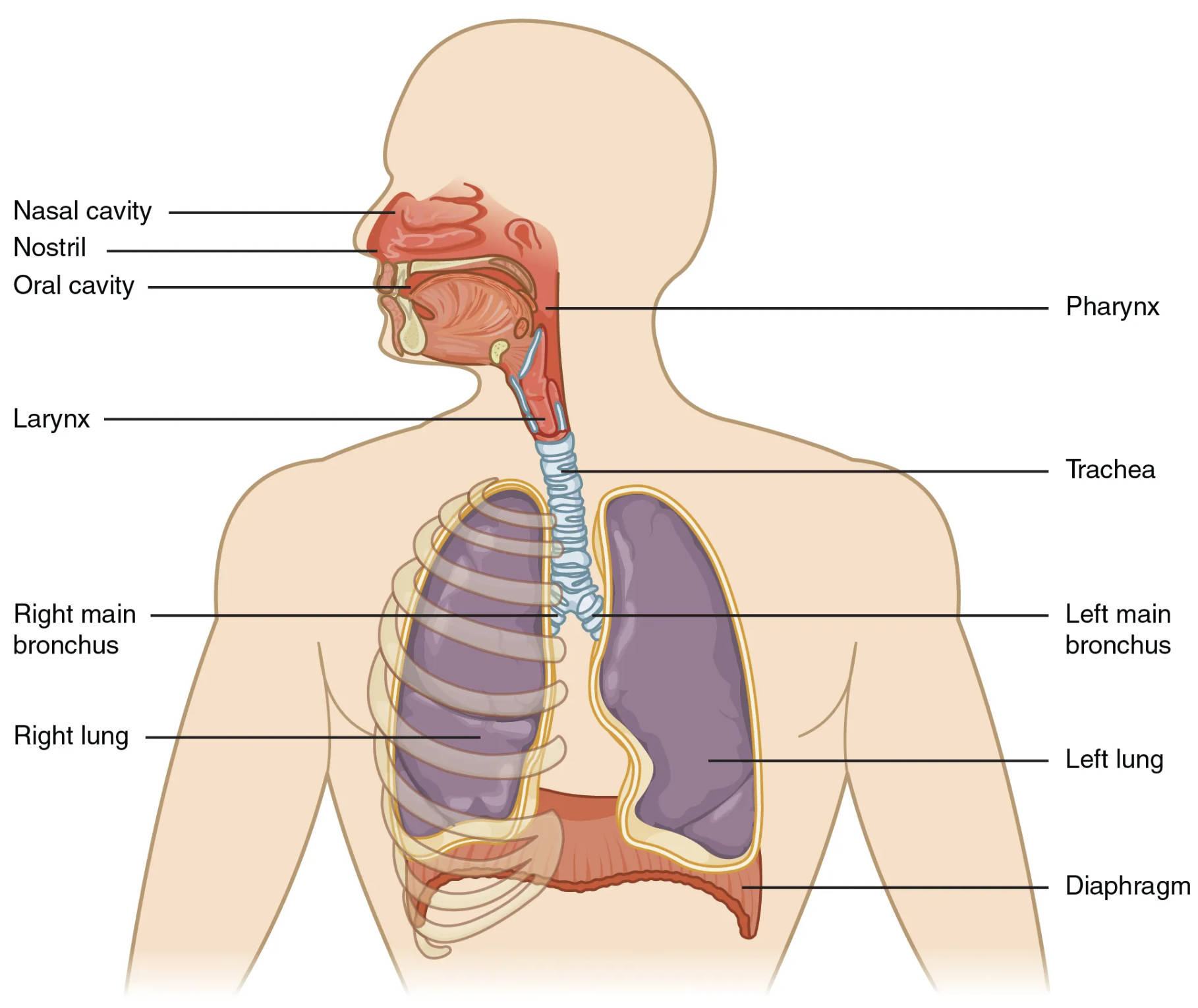
Each labeled part of the respiratory system plays a unique role in the process of breathing. These structures work together to facilitate air intake, filtration, and gas exchange.
Nasal cavity: The nasal cavity is the initial air entry point, where air is filtered, warmed, and moistened before moving deeper into the system. It contains mucous membranes and cilia that trap dust and pathogens, protecting the lower respiratory tract from infection.
Nostril: The nostril serves as the external opening to the nasal cavity, allowing air to enter and exit the respiratory system. It is lined with fine hairs that act as an additional filter, capturing larger particles before air reaches the nasal cavity.
Oral cavity: The oral cavity provides an alternative air entry route, particularly during heavy breathing or when the nasal passages are blocked. It connects to the pharynx and allows air to bypass the nasal filtration system in emergencies.
Pharynx: The pharynx, or throat, is a passageway that directs air from the nasal and oral cavities to the larynx and food to the esophagus. It contains muscles and tissues that assist in swallowing and prevent food from entering the airway.
Larynx: The larynx, known as the voice box, houses the vocal cords and produces sound as air passes through. It also acts as a protective sphincter, closing during swallowing to prevent aspiration into the lungs.
Trachea: The trachea is a sturdy tube supported by C-shaped cartilage rings, ensuring it remains open for air passage to the bronchi. Its inner lining, equipped with cilia and mucus, traps and removes debris, safeguarding the lungs.
Right main bronchus: The right main bronchus is the larger of the two primary bronchi, branching off the trachea to deliver air to the right lung. It contains cartilage and smooth muscle, allowing it to adjust its diameter during breathing.
Left main bronchus: The left main bronchus is slightly narrower and longer, extending from the trachea to the left lung. It adapts its structure to accommodate the heart’s position on the left side of the chest.
Right lung: The right lung, with three lobes, is the larger of the two lungs and handles a significant portion of gas exchange. It contains millions of alveoli where oxygen enters the bloodstream and carbon dioxide is expelled.
Left lung: The left lung, with two lobes, is slightly smaller to accommodate the heart and performs similar gas exchange functions as the right lung. Its structure supports efficient respiration despite its reduced size.
Diaphragm: The diaphragm is a dome-shaped muscle that separates the thoracic and abdominal cavities, playing a key role in breathing. It contracts to expand the chest cavity during inhalation and relaxes to aid exhalation.
Functions and Physiological Roles
The respiratory system’s structures contribute to vital physiological processes. This coordination ensures the body’s oxygen needs are met efficiently.
- Gas exchange occurs in the alveoli, where oxygen diffuses into the blood and carbon dioxide is removed, maintained by a pressure gradient.
- The system regulates blood pH by controlling carbon dioxide levels, which influence the formation of carbonic acid.
- Breathing is driven by the diaphragm and intercostal muscles, with the nervous system adjusting rate and depth based on oxygen demand.
- The larynx enables phonation, allowing sound production through vocal cord vibration.
- Mucous membranes and cilia provide a first line of defense against pathogens and pollutants.
Anatomical Interactions with Other Systems
The respiratory system collaborates with other bodily systems for optimal function. This interplay supports overall health and adaptability.
- It integrates with the cardiovascular system, delivering oxygenated blood via the pulmonary arteries and veins.
- The nervous system monitors oxygen and carbon dioxide levels, sending signals to adjust breathing via the medulla oblongata.
- Hormones like adrenaline from the endocrine system can enhance respiratory rate during stress or exercise.
- The immune system uses lymphatic tissue in the pharynx to combat respiratory infections.
- The skeletal system, particularly the ribs, provides structural support for lung expansion.
Maintaining Respiratory Health
Healthy respiratory function relies on lifestyle choices and awareness. These practices can prevent complications and enhance well-being.
- Avoiding tobacco smoke preserves lung tissue and prevents chronic obstructive pulmonary disease.
- Regular aerobic exercise strengthens the diaphragm and increases lung capacity.
- Staying hydrated keeps mucus thin, aiding in the removal of trapped particles.
- Using air purifiers in polluted environments reduces inhalation of harmful substances.
- Annual check-ups can detect early signs of respiratory issues, ensuring timely intervention.
In conclusion, the major structures of the respiratory system form a remarkable network that sustains life through efficient gas exchange and protection. By understanding how the nasal cavity, trachea, lungs, and diaphragm work together, individuals can appreciate the importance of maintaining this system. Adopting healthy habits and staying informed about its functions can lead to a lifetime of robust respiratory health, supporting all aspects of daily life.

