The brain’s intricate structure is revealed in this frontal section, showcasing key components of the basal nuclei and surrounding features. This image highlights the striatum with its subdivisions, the globus pallidus, lateral ventricle, and corpus callosum, providing a clear view of their spatial relationships within the cerebral cortex. Understanding these elements is essential for grasping motor control, cognitive functions, and overall neurological health.
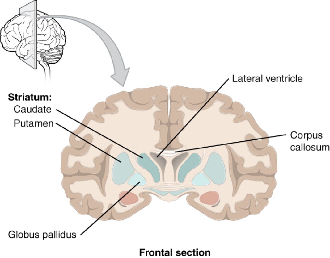
Striatum: Caudate
The caudate nucleus, part of the striatum, is located just lateral to the lateral ventricle and forms a C-shaped structure that extends into the temporal lobe. It plays a crucial role in motor planning, procedural learning, and cognitive processes, integrating inputs from the cerebral cortex and thalamus.
Striatum: Putamen
The putamen, another component of the striatum, lies inferior to the caudate and is separated by the internal capsule, a major white matter tract. This nucleus is involved in regulating movements and influencing various aspects of motor and sensory functions through its connections with the globus pallidus and substantia nigra.
Globus pallidus
The globus pallidus, positioned medial to the putamen, consists of internal and external segments that modulate motor activity via inhibitory projections. It acts as an output nucleus of the basal ganglia, fine-tuning voluntary movements and contributing to the direct and indirect pathways that control thalamic activity.
Lateral ventricle
The lateral ventricle is a C-shaped cavity filled with cerebrospinal fluid, located in each cerebral hemisphere and playing a key role in cushioning the brain and maintaining intracranial pressure. It connects to the third ventricle via the interventricular foramen, facilitating the circulation of cerebrospinal fluid produced by the choroid plexus.
Corpus callosum
The corpus callosum is a thick bundle of nerve fibers connecting the left and right cerebral hemispheres, enabling interhemispheric communication. This structure supports the integration of sensory, motor, and cognitive information, ensuring coordinated brain function across both sides.
Anatomical Overview of the Basal Nuclei
The basal nuclei, also known as basal ganglia, are subcortical structures critical for motor control and behavioral regulation. Their positioning in the frontal section illustrates their deep integration within the brain’s architecture.
- The basal nuclei include the striatum (caudate and putamen), globus pallidus, substantia nigra, and subthalamic nucleus, forming loops with the cortex and thalamus.
- These structures receive dopaminergic inputs from the substantia nigra, modulating pathways that either facilitate or inhibit movement.
- The internal capsule separates the caudate and putamen, carrying corticospinal and thalamocortical fibers essential for motor and sensory relay.
- Blood supply primarily comes from the lenticulostriate arteries, branches of the middle cerebral artery, ensuring metabolic support for high neuronal activity.
Detailed Structure of the Striatum
The striatum serves as the input station for the basal ganglia, processing cortical information. Its subdivisions, caudate and putamen, exhibit distinct yet complementary roles in neural circuits.
- The caudate nucleus wraps around the lateral ventricle, with its head in the frontal lobe, body in the parietal, and tail extending to the temporal lobe near the amygdala.
- The putamen, lens-shaped, forms the lentiform nucleus with the globus pallidus, involved in habit formation and reward-based learning via dopamine receptors.
- Striatal neurons are primarily medium spiny neurons, which are GABAergic and express D1 or D2 receptors, differentiating direct and indirect pathways.
- Connectivity includes projections to the globus pallidus and substantia nigra, influencing motor output through thalamic disinhibition.
Role and Anatomy of the Globus Pallidus
The globus pallidus functions as a regulatory hub within the basal ganglia. Its medial position relative to the putamen underscores its role in output modulation.
- Divided into globus pallidus externa (GPe) and interna (GPi), it receives inhibitory inputs from the striatum and sends outputs to the thalamus and subthalamic nucleus.
- The GPi projects to the ventrolateral thalamus, inhibiting unwanted movements, while GPe modulates the subthalamic nucleus in the indirect pathway.
- Composed mainly of large GABAergic neurons, it exhibits low spontaneous activity, increasing during movement suppression.
- Vascular supply from the anterior choroidal artery supports its function, with disruptions potentially leading to movement abnormalities.
Significance of the Lateral Ventricle
The lateral ventricle is a vital component of the ventricular system, providing structural and protective support. Its prominence in the frontal section highlights its proximity to basal structures.
- Each lateral ventricle consists of anterior, body, posterior, and inferior horns, containing choroid plexus that secretes cerebrospinal fluid at a rate of about 500 ml per day.
- Cerebrospinal fluid circulates through the ventricles, absorbing nutrients and removing waste, while maintaining buoyancy to reduce brain weight by 97%.
- The septum pellucidum separates the two lateral ventricles medially, and ependymal cells line the walls, facilitating fluid dynamics.
- Enlargement of the lateral ventricle, known as ventriculomegaly, can indicate atrophy or increased pressure, assessed via imaging.
Function of the Corpus Callosum
The corpus callosum facilitates bilateral brain coordination, visible as a central white matter tract in this section. Its role extends beyond mere connection to enabling complex cognitive integration.
- Comprising over 200 million axons, it is divided into rostrum, genu, body, and splenium, with fibers crossing to contralateral hemispheres.
- Anterior portions connect frontal lobes for executive functions, while posterior parts link occipital lobes for visual processing.
- Myelinated axons ensure rapid signal transmission, with conduction speeds up to 100 m/s, supporting tasks like bimanual coordination.
- Developmental anomalies, such as agenesis, can affect cognitive symmetry, studied through diffusion tensor imaging.
Integrated Functions and Neural Pathways
The interplay between these structures forms the foundation of motor and cognitive circuits. This frontal view emphasizes their collective contribution to brain physiology.
- Basal nuclei loops, including cortico-striato-pallido-thalamo-cortical pathways, regulate initiation and execution of movements.
- Dopamine from the substantia nigra modulates striatal activity, with D1 receptors exciting the direct pathway and D2 inhibiting the indirect.
- The corpus callosum integrates hemispheric inputs, while ventricles provide a fluid environment that dampens mechanical stress.
- Neurotransmitters like GABA in the globus pallidus and glutamate in cortical projections maintain balance in these circuits.
Clinical and Physiological Implications
Knowledge of this anatomy informs physiological understanding and clinical approaches. The structures’ vulnerabilities highlight their importance in maintaining homeostasis.
- The striatum’s role in reward processing involves dopamine release, influencing motivation and learning through mesolimbic pathways.
- Globus pallidus hyperactivity can disrupt motor control, as seen in dystonia, where GABAergic imbalance prevails.
- Ventricular system dynamics, regulated by aquaporin-4 channels, affect fluid resorption and pressure gradients.
- Corpus callosum integrity is crucial for split-brain studies, revealing lateralized functions like language in the left hemisphere.
Conclusion
This frontal section of the cerebral cortex and basal nuclei offers a profound insight into the brain’s subcortical architecture, with the striatum, globus pallidus, lateral ventricle, and corpus callosum each contributing uniquely to neural function. These components work in harmony to support movement, cognition, and protection, underscoring the brain’s sophisticated design. A thorough grasp of their anatomy enhances appreciation for neurological processes and aids in advancing medical knowledge.

