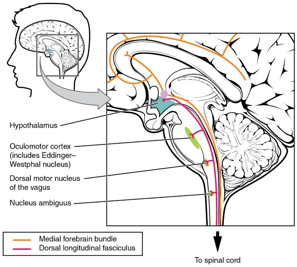
The fiber tracts of the central autonomic system diagram reveals the intricate network that governs the body’s involuntary functions, with the hypothalamus serving as the central hub. This chart illustrates how neural pathways, including the medial forebrain bundle and dorsal longitudinal fasciculus, connect the hypothalamus to the brainstem and spinal cord, regulating the balance between sympathetic and parasympathetic activities. Understanding these pathways provides a foundation for comprehending how the autonomic nervous system maintains homeostasis across various organ systems.
Labeled Components in the Diagram
Hypothalamus The hypothalamus acts as the primary control center for autonomic functions, integrating inputs from higher brain regions. It projects signals through specific tracts to regulate processes like heart rate and digestion.
Oculomotor cortex (includes Edinger-Westphal nucleus) The oculomotor cortex (includes Edinger-Westphal nucleus) is involved in coordinating eye movements and pupillary reflexes, with the Edinger-Westphal nucleus managing parasympathetic control. This region receives hypothalamic input to adjust pupil size in response to light.
Dorsal motor nucleus of the vagus The dorsal motor nucleus of the vagus is located in the medulla and controls parasympathetic output to the heart, lungs, and digestive tract via the vagus nerve. It receives regulatory signals from the hypothalamus to maintain visceral functions.
Nucleus ambiguus The nucleus ambiguus, also in the medulla, contributes to parasympathetic control of the heart and other organs through the vagus nerve. It modulates cardiac activity based on hypothalamic directives.
Medial forebrain bundle The medial forebrain bundle is a major tract connecting the hypothalamus to the brainstem and spinal cord, facilitating sympathetic and parasympathetic coordination. This pathway supports a wide range of autonomic responses, including temperature regulation.
Dorsal longitudinal fasciculus The dorsal longitudinal fasciculus runs along the brainstem, linking the hypothalamus to the dorsal vagal complex and other autonomic centers. It plays a key role in transmitting signals for visceral control and emotional responses.
To spinal cord The to spinal cord indicates the continuation of hypothalamic projections to spinal autonomic neurons. This connection allows for the regulation of sympathetic outflow to peripheral organs.
Anatomy of the Autonomic Pathways
The central autonomic system orchestrates involuntary bodily functions through a complex network of neural tracts. These pathways ensure seamless communication between the brain and peripheral organs.
- The hypothalamus integrates sensory and hormonal inputs to direct autonomic activity.
- The medial forebrain bundle serves as a bidirectional conduit for hypothalamic signals.
- The dorsal longitudinal fasciculus connects the hypothalamus to medullary centers.
- The oculomotor cortex and Edinger-Westphal nucleus regulate ocular reflexes.
- The dorsal motor nucleus of the vagus and nucleus ambiguus control parasympathetic functions.
- Spinal projections influence sympathetic ganglia for fight-or-flight responses.
Physiological Mechanisms of Autonomic Control
The physiological processes governed by these tracts involve neurotransmitter release and neural integration. This ensures precise regulation of bodily functions in response to internal and external cues.
- The hypothalamus releases corticotropin-releasing hormone to modulate stress responses.
- The medial forebrain bundle transmits dopamine and serotonin for emotional regulation.
- The dorsal longitudinal fasciculus carries signals for vagal tone adjustment.
- Acetylcholine from the Edinger-Westphal nucleus constricts pupils via parasympathetic action.
- The dorsal motor nucleus of the vagus secretes acetylcholine to slow heart rate.
- Sympathetic activation via spinal pathways releases norepinephrine.
Role of the Hypothalamus
The hypothalamus is the master regulator of the autonomic nervous system. Its strategic location and connectivity underpin its critical role.
- It receives inputs from the limbic system to influence emotional-driven responses.
- The suprachiasmatic nucleus within the hypothalamus controls circadian rhythms.
- It projects to the brainstem via the medial forebrain bundle and dorsal longitudinal fasciculus.
- The paraventricular nucleus releases oxytocin and vasopressin for homeostasis.
- It modulates body temperature through sympathetic pathways to the spinal cord.
- Damage to the hypothalamus can disrupt autonomic balance, affecting vital functions.
Clinical Relevance of Autonomic Tracts
Understanding these fiber tracts aids in diagnosing and treating disorders of the autonomic nervous system. Their assessment provides insights into patient health.
- Lesions in the medial forebrain bundle may cause appetite or sleep disturbances.
- Dysfunction in the dorsal longitudinal fasciculus can lead to vagal nerve issues.
- Edinger-Westphal nucleus impairment results in abnormal pupillary responses.
- Dorsal motor nucleus of the vagus damage may cause gastrointestinal motility issues.
- Spinal cord injuries disrupt sympathetic control, leading to orthostatic hypotension.
- Neuroimaging helps locate tract abnormalities in autonomic dysfunction.
Interaction with Peripheral Systems
The central autonomic tracts interface with peripheral ganglia to execute bodily functions. This interaction ensures coordinated organ responses.
- The nucleus ambiguus influences heart rate via the vagus nerve’s cardiac branches.
- The dorsal motor nucleus of the vagus regulates gastric secretion and motility.
- Sympathetic preganglionic neurons from the spinal cord synapse in paravertebral ganglia.
- The medial forebrain bundle connects to the sympathetic chain for stress responses.
- Parasympathetic outflow adjusts pupil size and salivary gland activity.
- These pathways maintain a balance between energy conservation and expenditure.
Advances in Autonomic Research
Recent studies enhance our knowledge of autonomic fiber tracts and their therapeutic potential. These developments offer hope for improved treatments.
- Functional MRI maps hypothalamic connectivity to autonomic centers.
- Deep brain stimulation targets the medial forebrain bundle for depression.
- Gene therapy aims to repair dorsal longitudinal fasciculus damage.
- Pharmacological agents modulate hypothalamic hormone release.
- Optogenetics traces neural pathways in animal models.
- Wearable sensors monitor autonomic responses in real-time.
The fiber tracts of the central autonomic system highlight the brain’s sophisticated control over involuntary functions, from heart rate to digestion. Mastering these pathways equips professionals to address a range of autonomic disorders, paving the way for innovative treatments and better patient care through ongoing research and clinical application.

