The female reproductive system is a marvel of biological engineering, responsible for reproduction, hormone production, and supporting pregnancy. This detailed guide, informed by anatomical views, delves into the intricate structures that comprise this vital system. Understanding its components, from external genitalia to internal organs, is crucial for appreciating female health and physiology.
https://anatomynote.com/pictures/reproductive-system/001/female-reproductive-system-anterior-and-lateral-view.jpg
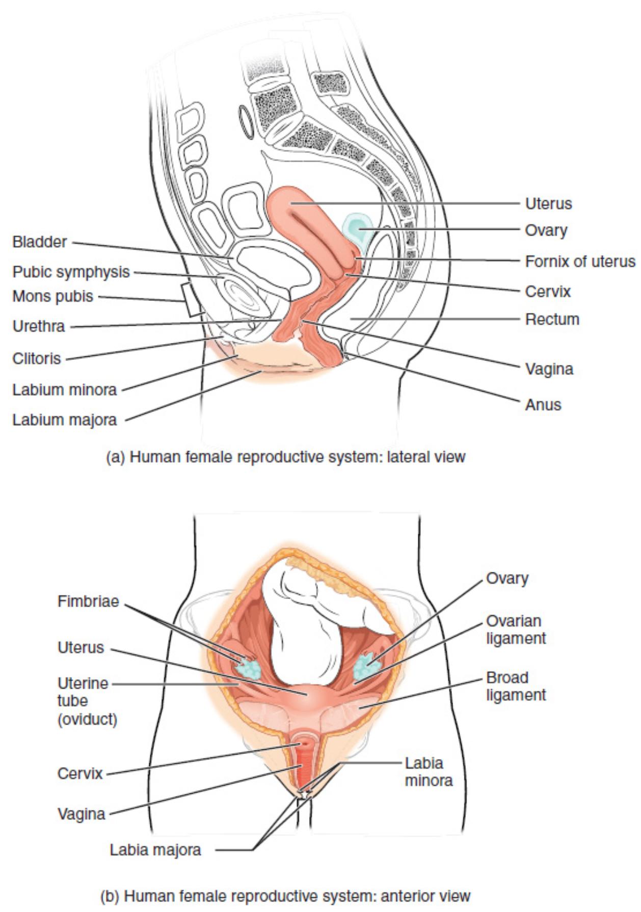
Uterus: The uterus is a hollow, pear-shaped muscular organ located in the pelvic cavity, between the bladder and the rectum. Its primary function is to nurture the developing fetus during pregnancy, and it also plays a crucial role in menstruation.
Ovary: The ovaries are a pair of small, almond-shaped glands located on either side of the uterus. They are responsible for producing ova (eggs) and secreting essential female hormones like estrogen and progesterone.
Fornix of uterus: The fornix refers to the recessed areas around the cervix where it projects into the vagina. These anatomical pouches, anterior, posterior, and lateral, can be clinically significant during examinations and procedures.
Cervix: The cervix is the lower, narrow part of the uterus that forms a canal opening into the vagina. It acts as a gateway, allowing sperm to enter and menstrual blood to exit, and it dilates during childbirth.
Rectum: The rectum is the final section of the large intestine, terminating at the anus. While not part of the reproductive system, its close anatomical proximity is important for understanding pelvic organ relationships and potential clinical interactions.
Vagina: The vagina is a muscular, elastic tube that connects the uterus to the outside of the body. It serves as the birth canal, the receptacle for sperm during intercourse, and the pathway for menstrual flow.
Anus: The anus is the external opening at the end of the gastrointestinal tract, through which feces are expelled. Similar to the rectum, its proximity to reproductive structures is noteworthy in pelvic anatomy.
Bladder: The bladder is a hollow, muscular organ located in the pelvic cavity that stores urine. Its anterior position relative to the uterus and vagina is a key anatomical landmark in the female pelvis.
Pubic symphysis: The pubic symphysis is a cartilaginous joint that connects the left and right pubic bones at the front of the pelvis. This joint provides flexibility to the pelvis, particularly important during childbirth.
Mons pubis: The mons pubis is a mound of fatty tissue located anterior to the pubic bone, typically covered with pubic hair after puberty. It serves as a protective cushion over the pubic symphysis.
Urethra: The urethra is a tube that transports urine from the bladder to the outside of the body. In females, it is shorter than in males and is located anterior to the vagina.
Clitoris: The clitoris is a small, highly sensitive erectile organ located at the anterior junction of the labia minora. It is rich in nerve endings and is a primary center for sexual sensation.
Labium minora: The labia minora are two delicate folds of skin located medial to the labia majora, enclosing the vestibule. These folds protect the clitoris and the openings of the urethra and vagina.
Labium majora: The labia majora are two prominent, fleshy folds of skin that form the outer boundaries of the vulva. They provide protection to the more delicate internal structures of the external genitalia.
Fimbriae: Fimbriae are finger-like projections located at the end of the fallopian (uterine) tubes, near the ovary. Their rhythmic sweeping motion helps to capture the ovum released from the ovary, guiding it into the uterine tube.
Uterine tube (oviduct): The uterine tubes, also known as fallopian tubes or oviducts, are a pair of muscular tubes extending from the uterus towards the ovaries. These tubes are the usual site of fertilization, transporting the ovum to the uterus.
Ovarian ligament: The ovarian ligament is a fibrous band that connects the medial pole of the ovary to the lateral wall of the uterus. It helps to anchor the ovary in its position within the pelvic cavity.
Broad ligament: The broad ligament is a wide fold of peritoneum that drapes over the uterus, uterine tubes, and ovaries. It helps to keep these organs in place within the pelvis and contains blood vessels and nerves.
Unveiling the Female Reproductive Anatomy
The female reproductive system is a marvel of biological design, intricately structured to facilitate reproduction, produce essential hormones, and support the development of new life. Comprising both external genitalia and internal organs, its complexity is fundamental to human perpetuation. The provided diagrams offer both a lateral and an anterior perspective, allowing for a comprehensive understanding of how these vital structures are positioned and interact within the pelvic cavity.
Understanding the anatomy of this system is not merely academic; it is crucial for comprehending reproductive health, contraception, fertility, and the various physiological changes a woman experiences throughout her life. Each component, from the egg-producing ovaries to the nurturing uterus, plays a distinct yet interconnected role in this biological symphony.
Key aspects of the female reproductive system include:
-
- The production of ova (eggs) and female hormones by the ovaries.
- The pathways for fertilization and implantation, involving the uterine tubes and uterus.
- The protective and sensory functions of the external genitalia.
- The supportive ligaments and peritoneal folds that hold organs in place.
Disruptions or conditions affecting any part of this system can have significant impacts on a woman’s health and reproductive capabilities, underscoring the importance of its intricate and coordinated functions.
Internal and External Structures
The internal female reproductive organs are primarily located within the pelvic cavity, nestled between the bladder anteriorly and the rectum posteriorly. The uterus, a central organ, is a muscular, hollow structure where a fertilized egg implants and develops. Its thick walls provide protection and nourishment to the growing fetus. Extending laterally from the uterus are the uterine tubes, or fallopian tubes, which feature fimbriae at their ovarian ends. These fimbriae gently sweep over the ovaries to capture the ovum released during ovulation, guiding it into the tube where fertilization typically occurs. The ovaries themselves are glandular organs, vital for both gamete production and hormone secretion, playing a crucial role in the menstrual cycle and the maintenance of female characteristics.
The external female genitalia, collectively known as the vulva, serve protective, sensory, and reproductive functions. These structures include the labia majora and minora, which are folds of skin and mucous membrane protecting the sensitive inner areas. The clitoris, a highly innervated erectile tissue, is central to sexual sensation. The urethral opening, for urine excretion, and the vaginal opening, for intercourse and childbirth, are also part of the vulva. The close anatomical relationship between the urinary and reproductive systems in females is evident in the arrangement of these external structures. Together, the internal and external components of the female reproductive system form a cohesive unit, essential for the continuation of the human species and for a woman’s overall health.
In conclusion, the female reproductive system is an exquisitely designed and functionally critical part of the human body. Its complex architecture, from the hormone-producing ovaries to the nurturing uterus and the protective external structures, is dedicated to reproduction and the delicate balance of female physiology. A thorough understanding of these anatomical relationships and their physiological roles is paramount for maintaining reproductive health, addressing related medical conditions, and appreciating the intricate processes of life.

