The extraocular muscles play a pivotal role in controlling eye movement and maintaining proper alignment within the orbit. This lateral view of the right eye provides a clear depiction of these muscles and their anatomical relationships, offering insight into their coordinated function.
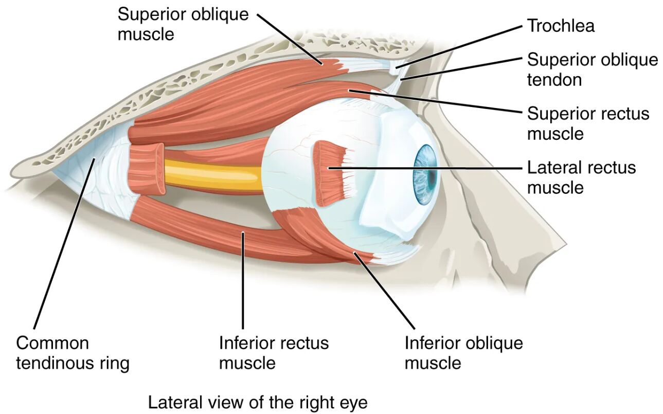
Superior oblique muscle The superior oblique muscle rotates the eye downward and outward, aiding in downward gaze and intorsion. It originates near the common tendinous ring and passes through the trochlea to exert its unique action.
Trochlea The trochlea is a cartilage structure that acts as a pulley, guiding the superior oblique tendon. This mechanism enhances the muscle’s ability to adjust the eye’s position with precision.
Superior oblique tendon The superior oblique tendon connects the superior oblique muscle to the eyeball, passing through the trochlea. It transmits the muscle’s force, enabling effective rotational movement.
Superior rectus muscle The superior rectus muscle elevates the eye and assists in inward rotation, crucial for upward gaze. It is innervated by the oculomotor nerve, ensuring smooth coordination with other muscles.
Lateral rectus muscle The lateral rectus muscle abducts the eye, moving it outward away from the nose. Controlled by the abducens nerve, it plays a key role in lateral eye movements.
Inferior rectus muscle The inferior rectus muscle depresses the eye and aids in outward rotation, facilitating downward gaze. It works in tandem with the superior rectus for vertical alignment.
Inferior oblique muscle The inferior oblique muscle elevates and outwardly rotates the eye, supporting upward and lateral movements. Its insertion pattern allows for versatile eye tracking.
Common tendinous ring The common tendinous ring is a fibrous structure where the rectus muscles originate, anchoring them to the orbit. It provides a stable foundation for coordinated eye motion.
Anatomy of the Extraocular Muscles
The extraocular muscles are a set of six muscles that surround the eyeball, depicted here in a lateral view of the right eye. Their arrangement and attachments are critical for maintaining visual alignment and movement.
- The superior and inferior rectus muscles handle vertical eye motion, with secondary rotational effects.
- The lateral rectus muscle specializes in abduction, balancing the medial rectus on the opposite side.
- The superior oblique muscle, aided by the trochlea, adds torsional control to downward movements.
- The inferior oblique muscle complements the superior oblique, enhancing upward and outward motion.
- The common tendinous ring, also called the annulus of Zinn, serves as the origin for the rectus muscles.
- Tendons, such as the superior oblique tendon, transmit force from muscles to the eyeball.
- The orbit’s bony structure protects these muscles while allowing their dynamic range of motion.
Functions of Eye Movement
Each extraocular muscle contributes to specific eye movements, enabling a wide range of visual tasks. Their synchronized action ensures stable gaze and accurate tracking.
- The superior rectus muscle lifts the eye, assisting in looking up or focusing on elevated objects.
- The lateral rectus muscle moves the eye outward, essential for peripheral vision or following motion.
- The inferior rectus muscle lowers the eye, aiding in downward gaze like reading or floor inspection.
- The superior oblique muscle, via the trochlea, rotates the eye downward and outward for fine adjustments.
- The inferior oblique muscle elevates and rotates the eye, supporting upward and lateral tracking.
- These muscles operate in pairs, with agonists and antagonists maintaining balance.
- Rapid eye movements, or saccades, rely on this muscular coordination for quick focus shifts.
- The system adapts to sustained positions, adjusting tension to prevent fatigue.
Role of the Trochlea and Tendons
The trochlea and tendons enhance the mechanical efficiency of the superior oblique muscle. This setup allows for intricate control over eye rotation and alignment.
- The trochlea redirects the superior oblique tendon, optimizing its pull angle for effective rotation.
- The superior oblique tendon, guided by the trochlea, reduces friction and wear during movement.
- This pulley system enables the muscle to exert torque, rotating the eye along its visual axis.
- Tendons are made of dense collagen, offering strength and flexibility for repeated use.
- The trochlea’s cartilage composition minimizes stress on the tendon, ensuring durability.
- Misalignment of the trochlea can affect superior oblique function, though this diagram shows normal anatomy.
Innervation and Coordination
The extraocular muscles are innervated by cranial nerves, ensuring precise and rapid responses. This neural control integrates with the brain for seamless eye movement.
- The oculomotor nerve (CN III) supplies the superior and inferior rectus muscles.
- The trochlear nerve (CN IV) innervates the superior oblique muscle, guiding its unique path.
- The abducens nerve (CN VI) controls the lateral rectus muscle, specializing in abduction.
- These nerves originate from the brainstem, with nuclei coordinating bilateral eye actions.
- Reflex arcs, like the vestibulo-ocular reflex, stabilize gaze during head motion.
- Damage to these nerves can lead to strabismus, but this image illustrates healthy function.
Clinical Relevance of Extraocular Muscles
Understanding these muscles aids in diagnosing and managing eye movement disorders, though this image depicts normal anatomy. Knowledge of their structure supports effective clinical approaches.
- Weakness in the lateral rectus muscle may cause esotropia, where the eye turns inward.
- Superior oblique dysfunction, linked to trochlea issues, can result in vertical diplopia.
- The inferior rectus muscle’s impairment might hinder downward gaze, affecting daily tasks.
- Muscle balance is assessed via tests like the cover test or eye movement recordings.
- Surgical techniques, such as muscle recession, correct alignment in strabismus cases.
- Rehabilitation exercises can strengthen these muscles after nerve palsy.
- Imaging and electromyography help evaluate muscle and nerve health.
In conclusion, the extraocular muscles, as shown in this lateral view, form a sophisticated system for eye movement and stability. Their precise coordination, supported by tendons and neural innervation, highlights the complexity of maintaining clear vision and spatial orientation.

