The heart, a vital organ encased within the pericardium, showcases a complex network of structures essential for sustaining life. This article delves into the external anatomy of the heart, presenting detailed views from both the anterior and posterior perspectives to highlight its major features and their functions. Understanding these components provides a foundation for appreciating the heart’s role in circulation and overall cardiovascular health.
Brachiocephalic trunk
- This major artery branches from the aortic arch, supplying blood to the right arm and head.
- It plays a critical role in delivering oxygenated blood to the upper body regions.
Superior vena cava
- The superior vena cava is a large vein that returns deoxygenated blood from the upper half of the body to the right atrium.
- Its position near the heart ensures efficient blood flow for oxygenation.
Right pulmonary artery
- This artery carries deoxygenated blood from the right ventricle to the lungs for oxygenation.
- Its connection to the pulmonary trunk facilitates the pulmonary circulation process.
Ascending aorta
- The ascending aorta rises from the left ventricle, distributing oxygenated blood to the systemic circulation.
- It serves as the initial segment of the aorta, critical for maintaining blood pressure.
Pulmonary trunk
- The pulmonary trunk transports deoxygenated blood from the right ventricle to the lungs via the pulmonary arteries.
- It divides into the left and right pulmonary arteries, marking the start of pulmonary circulation.
Right pulmonary veins
- These veins return oxygenated blood from the lungs to the left atrium.
- Their function is essential for maintaining the oxygen supply to the systemic circulation.
Right atrium
- The right atrium receives deoxygenated blood from the body via the superior and inferior vena cava.
- It acts as a chamber that pumps blood into the right ventricle.
Right coronary artery
- This artery supplies oxygenated blood to the right atrium, right ventricle, and parts of the left ventricle.
- It is vital for nourishing the heart muscle and preventing ischemic damage.
Anterior cardiac vein
- The anterior cardiac vein drains deoxygenated blood from the front of the heart into the right atrium.
- It supports the coronary circulation by removing metabolic waste.
Right ventricle
- The right ventricle pumps deoxygenated blood into the pulmonary trunk for lung oxygenation.
- Its muscular walls are adapted to handle lower pressure compared to the left ventricle.
Right marginal artery
- This artery branches from the right coronary artery, supplying blood to the right ventricle’s margin.
- It ensures adequate perfusion to the heart’s lateral regions.
Small cardiac vein
- The small cardiac vein drains deoxygenated blood from the posterior heart surface into the coronary sinus.
- It contributes to the heart’s venous return system.
Inferior vena cava
- The inferior vena cava returns deoxygenated blood from the lower body to the right atrium.
- Its large diameter accommodates significant blood volume from the legs and abdomen.
Left common carotid artery
- This artery supplies oxygenated blood to the head and neck, branching from the aortic arch.
- It is crucial for cerebral circulation and brain function.
Left subclavian artery
- The left subclavian artery provides blood to the left arm and parts of the brain.
- It originates from the aortic arch, ensuring upper limb perfusion.
Aortic arch
- The aortic arch curves between the ascending and descending aorta, distributing blood to the upper body.
- It gives rise to major arteries like the brachiocephalic trunk and left carotid artery.
Ligamentum arteriosum
- This ligament is a remnant of the ductus arteriosus, connecting the pulmonary artery to the aorta in fetal life.
- It becomes fibrous after birth, marking a key developmental transition.
Left pulmonary artery
- The left pulmonary artery carries deoxygenated blood from the right ventricle to the left lung.
- It works in tandem with the right pulmonary artery to support lung oxygenation.
Left pulmonary veins
- These veins return oxygenated blood from the left lung to the left atrium.
- They are essential for delivering oxygen-rich blood to the systemic circulation.
Auricle of left atrium
- The auricle is a small, ear-like extension of the left atrium that increases its volume.
- It aids in receiving blood from the pulmonary veins.
Circumflex artery
- The circumflex artery, a branch of the left coronary artery, supplies blood to the left atrium and ventricle.
- It is critical for preventing myocardial ischemia in the posterior heart regions.
Left coronary artery
- This artery originates from the aortic root, supplying blood to the left atrium and ventricle.
- It plays a key role in nourishing the heart’s left side.
Left ventricle
- The left ventricle pumps oxygenated blood into the aorta for systemic circulation.
- Its thick, muscular walls generate high pressure to support body-wide blood flow.
Great cardiac vein
- The great cardiac vein drains deoxygenated blood from the anterior heart into the coronary sinus.
- It is a major component of the heart’s venous drainage system.
Anterior interventricular artery
- This artery, also called the left anterior descending artery, supplies blood to the interventricular septum and front of the left ventricle.
- It is vital for preventing anterior wall myocardial infarction.
Apex
- The apex is the pointed lower end of the heart, resting on the diaphragm.
- It marks the heart’s base-to-apex orientation and aids in its positioning.
Coronary sinus
- The coronary sinus collects deoxygenated blood from the heart’s veins and empties it into the right atrium.
- It serves as a central drainage point for coronary circulation.
Posterior vein of left ventricle
- This vein drains deoxygenated blood from the posterior left ventricle into the coronary sinus.
- It supports the venous return from the heart’s posterior regions.
Middle cardiac vein
- The middle cardiac vein drains the posterior interventricular septum into the coronary sinus.
- It ensures proper venous drainage from the heart’s lower regions.
Posterior interventricular artery
- This artery supplies blood to the posterior interventricular septum and portions of both ventricles.
- It is a branch of the right coronary artery in most individuals.
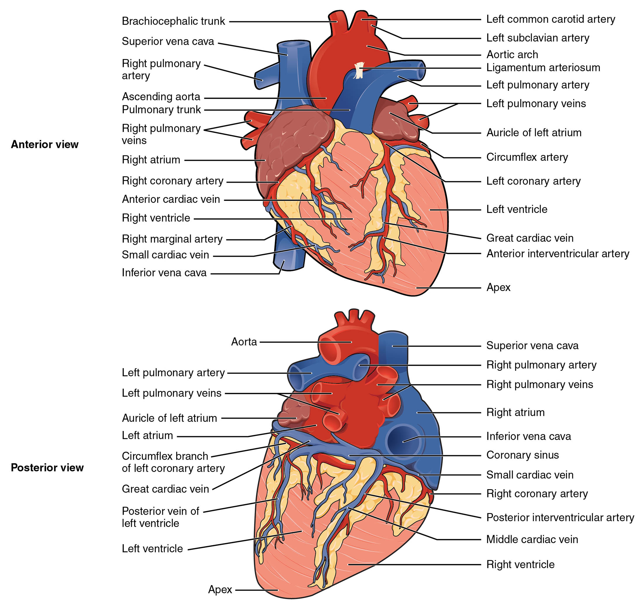
The heart’s external anatomy is a testament to its role as the powerhouse of the circulatory system. The anterior and posterior views reveal a sophisticated arrangement of arteries, veins, and chambers that work together to pump blood effectively. This exploration covers the key structures visible on the heart’s surface, providing a comprehensive look at its external features.
Anterior View: Unveiling the Heart’s Frontal Structure
The anterior view of the heart offers a clear perspective on the structures visible from the front. This view highlights the major vessels and chambers that initiate the journey of blood through the body.
- The ascending aorta and pulmonary trunk are prominently displayed, marking the outflow tracts of the left and right ventricles.
- Key arteries like the right coronary artery and anterior cardiac vein illustrate the heart’s self-sustaining blood supply.
Posterior View: Exploring the Heart’s Backside
The posterior view provides insight into the heart’s less visible but equally important structures. This perspective emphasizes the venous return and posterior arterial supply.
- The coronary sinus and posterior interventricular artery are central to this view, managing blood drainage and supply to the back of the heart.
- Veins such as the great cardiac vein and middle cardiac vein underscore the heart’s complex venous network.
The heart’s external anatomy is a critical area of study for understanding its function within the circulatory system. Encased in the pericardium, the heart’s surface features include a network of arteries and veins that ensure its continuous operation. The anterior view showcases the ascending aorta, pulmonary trunk, and right coronary artery, which are essential for distributing oxygenated and deoxygenated blood. The right atrium and ventricle, visible from this angle, receive and pump blood, respectively, setting the stage for pulmonary and systemic circulation.
Turning to the posterior view, structures like the coronary sinus, left pulmonary veins, and posterior interventricular artery come into focus. These components are vital for returning blood to the heart and supplying the posterior regions with oxygen-rich blood. The left atrium, receiving oxygenated blood from the lungs, and the left ventricle, pumping it into the aorta, demonstrate the heart’s dual circulatory roles. The apex, the heart’s lowest point, anchors its position within the thoracic cavity.
Arteries such as the brachiocephalic trunk and left common carotid artery branch from the aortic arch, supplying the head, neck, and upper limbs. The pulmonary arteries and veins facilitate the exchange of gases in the lungs, a process dependent on the heart’s rhythmic contractions. Veins like the superior and inferior vena cava return deoxygenated blood to the right atrium, completing the circulatory loop. The ligamentum arteriosum, a remnant of fetal circulation, adds a historical layer to the heart’s anatomical story.
The heart’s muscular walls, particularly the thick left ventricle, are adapted to handle the high pressure required for systemic circulation. The right ventricle, with thinner walls, manages the lower pressure of pulmonary circulation. The auricles of the atria enhance their capacity to receive blood, while the coronary arteries—right, left, and their branches like the circumflex and anterior interventricular—ensure the heart muscle itself is nourished. The great cardiac vein and its counterparts drain metabolic waste, maintaining the heart’s efficiency.
This anatomical overview highlights the heart’s resilience and complexity. The interplay of its external structures supports its role as a pump, driving blood through the pulmonary and systemic circuits. For those delving into cardiovascular physiology, recognizing these features provides a solid basis for understanding heart function and potential clinical implications.

