The human brain stands as a remarkable organ, with the cerebrum representing its largest and most intricate component. This anterior view of the cerebrum showcases key structures such as the right hemisphere, left hemisphere, and longitudinal fissure, providing valuable insights into the brain’s symmetry and functional organization within the central nervous system (CNS). This article delves into the anatomy and significance of these features, offering a comprehensive guide to understanding their roles in health and cognition.
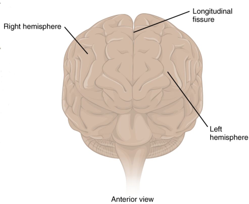
Right hemisphere
The right hemisphere of the cerebrum controls the left side of the body and is predominantly involved in spatial abilities, facial recognition, and processing visual and auditory stimuli. This half of the brain works in concert with the left hemisphere to ensure balanced sensory and motor functions.
Left hemisphere
The left hemisphere governs the right side of the body and is primarily responsible for language processing, logical reasoning, and analytical tasks. It collaborates with the right hemisphere through the corpus callosum to integrate diverse cognitive functions effectively.
Longitudinal fissure
The longitudinal fissure is a deep groove that separates the right and left hemispheres of the cerebrum. This structure houses the falx cerebri, a fold of dura mater, and plays a crucial role in maintaining the brain’s structural integrity while allowing interhemispheric communication.
Anatomical Overview of the Cerebrum
The cerebrum is the powerhouse of the brain, driving complex functions essential for daily life. Its anterior view reveals the symmetrical division that underpins its operational efficiency.
- The cerebrum constitutes about 85% of the brain’s total weight, highlighting its dominance in neural activity.
- It is divided into the right and left hemispheres by the longitudinal fissure, each containing four lobes: frontal, parietal, temporal, and occipital.
- The cerebral cortex, with its characteristic folds or gyri, covers the surface and enhances the brain’s computational capacity.
- Blood flow to the cerebrum, supplied by the anterior and middle cerebral arteries, is vital for sustaining its metabolic demands.
Detailed Analysis of the Hemispheres
The right and left hemispheres each contribute uniquely to brain function, creating a balanced system. Their distinct roles are critical for overall neurological health.
- The right hemisphere excels in nonverbal tasks, such as interpreting emotions from facial expressions and navigating spatial environments.
- The left hemisphere is dominant for language in most individuals, housing areas like Broca’s and Wernicke’s regions for speech production and comprehension.
- Both hemispheres process sensory input from the opposite side of the body, a phenomenon known as contralateral control.
- Hemispheric specialization can vary, with some individuals showing mixed dominance, particularly in creative or musical abilities.
Understanding the Longitudinal Fissure
The longitudinal fissure serves as a physical and functional divider, yet it supports connectivity between hemispheres. This structure is integral to the brain’s organizational framework.
- The fissure extends from the front of the brain to the back, creating a clear boundary that is bridged by the corpus callosum.
- It contains the falx cerebri, which provides structural support and prevents excessive lateral movement of the hemispheres.
- Surgical procedures, such as corpus callosotomy, may involve this region to treat severe epilepsy by limiting seizure spread.
- The depth and width of the fissure can vary, influencing the degree of interhemispheric interaction.
Clinical Significance and Functional Roles
The anterior view of the cerebrum offers insights into its clinical relevance, aiding in the diagnosis and treatment of neurological conditions. These structures are key to understanding brain pathology.
- Hemispheric damage, such as from a stroke, can lead to hemiplegia or aphasia, depending on the affected side and region.
- The longitudinal fissure’s proximity to the corpus callosum makes it a focal point in studies of interhemispheric disconnection syndromes.
- Imaging techniques like CT scans or MRI can assess hemisphere integrity, guiding interventions for traumatic brain injuries.
- Research into hemispheric dominance continues to inform therapies for conditions like dyslexia or spatial neglect.
Conclusion
The anterior view of the cerebrum provides a window into the brain’s symmetrical yet specialized architecture, with the right hemisphere, left hemisphere, and longitudinal fissure playing essential roles. These structures underpin the brain’s ability to process information, coordinate movements, and maintain cognitive harmony. A deeper understanding of their anatomy enhances our ability to address neurological challenges and appreciate the brain’s remarkable complexity.

