The alpha-beta T cell receptor (TCR) is a critical component of the adaptive immune system, enabling T cells to recognize and respond to specific antigens presented by major histocompatibility complex (MHC) molecules. Anchored within the T cell membrane, this receptor features distinct constant and variable regions that allow for precise antigen recognition and immune activation. This detailed illustration highlights the structural elements of the alpha-beta T cell receptor, offering a deeper understanding of its role in immunity.
Labeled Components of the Alpha-Beta T Cell Receptor
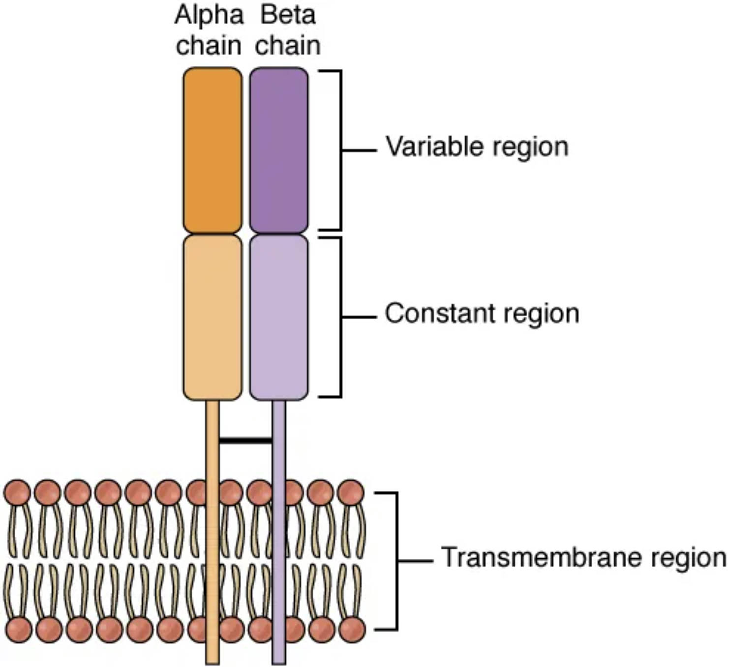
Alpha chain: This polypeptide forms one half of the TCR, containing a variable region that binds to antigens and a constant region for structural stability. It pairs with the beta chain to create the antigen-binding site.
Beta chain: The other half of the TCR, this chain also has variable and constant regions, contributing to antigen specificity. It works in tandem with the alpha chain to recognize MHC-presented peptides.
Variable region (alpha): Located at the tip of the alpha chain, this region exhibits diversity to recognize specific antigens. Its unique sequence allows the TCR to adapt to a wide range of pathogens.
Variable region (beta): Found on the beta chain, this region complements the alpha chain’s variable region in antigen recognition. It enhances the TCR’s ability to detect diverse MHC-antigen complexes.
Constant region (alpha): This stable segment of the alpha chain anchors the receptor to the T cell membrane and interacts with co-receptors. It ensures proper positioning and signaling within the immune response.
Constant region (beta): Positioned on the beta chain, this region provides structural support and connects to intracellular signaling molecules. It facilitates the transmission of activation signals.
Transmembrane region: This hydrophobic segment spans the T cell membrane, anchoring the TCR to the cell surface. It links the extracellular receptor to intracellular signaling pathways.
Intracellular region: Located inside the T cell, this region interacts with signaling molecules like CD3 to initiate immune activation. It relays the antigen recognition signal to trigger T cell responses.
Antigen-MHC complex: This structure, formed by an antigen peptide bound to an MHC molecule, is the target recognized by the TCR. It is presented on the surface of antigen-presenting cells to activate T cells.
Anatomical Structure of the Alpha-Beta T Cell Receptor
The alpha-beta TCR’s design is tailored for precise antigen recognition and immune signaling.
- The alpha and beta chains form a heterodimer, creating a functional receptor unit.
- Variable regions on both chains allow for extensive diversity, enabling recognition of countless antigens.
- Constant regions provide stability, anchoring the receptor within the membrane.
- The transmembrane region ensures the TCR remains embedded in the T cell surface.
- The intracellular region connects to co-receptors, amplifying the immune signal.
- The antigen-MHC complex serves as the specific ligand, triggering T cell activation.
This illustration emphasizes the receptor’s structural complexity and adaptability.
Physiological Role in Immune Function
The alpha-beta TCR plays a pivotal role in coordinating T cell-mediated immunity.
- The variable regions bind to the antigen-MHC complex, initiating T cell recognition.
- The constant regions stabilize the receptor, ensuring effective antigen engagement.
- The transmembrane region anchors the TCR, facilitating signal transduction.
- The intracellular region activates pathways involving CD3 and zeta chains.
- This recognition process activates cytotoxic or helper T cells, depending on the context.
- The receptor’s diversity allows responses to both viral and bacterial threats.
This function underscores its importance in adaptive immunity.
Molecular Interactions and Signaling
The TCR’s interactions with other molecules enhance its immune effectiveness.
- The alpha and beta chains’ variable regions undergo V(D)J recombination for diversity.
- The constant regions associate with CD3 complex, amplifying signal strength.
- The transmembrane region integrates with the lipid bilayer, stabilizing receptor placement.
- The intracellular region recruits kinases like Lck to initiate signaling cascades.
- The antigen-MHC complex binding triggers calcium influx and gene expression.
- Co-receptors like CD4 or CD8 enhance affinity, boosting T cell activation.
These interactions ensure a robust and specific immune response.
Clinical Relevance of the Alpha-Beta T Cell Receptor
Understanding the TCR’s structure aids in managing immune-related conditions.
- Defects in variable region diversity can lead to immunodeficiency, reducing antigen recognition.
- Constant region mutations may impair signaling, affecting T cell function.
- Transmembrane region issues can disrupt receptor anchoring, weakening immunity.
- Intracellular signaling defects are linked to autoimmune diseases like multiple sclerosis.
- Altered antigen-MHC binding is studied in transplant rejection and cancer immunotherapy.
- TCR analysis helps develop targeted therapies, such as CAR-T cell treatments.
This knowledge is vital for advancing immunological treatments.
Developmental and Adaptive Features
The TCR adapts throughout life, reflecting its dynamic immune role.
- The alpha and beta chains undergo gene rearrangement during T cell development.
- Variable regions diversify in the thymus, ensuring broad antigen coverage.
- Constant regions remain stable, supporting consistent signaling across T cell types.
- The transmembrane region matures with the T cell, ensuring proper membrane integration.
- The intracellular region evolves to optimize signal transduction efficiency.
- Antigen-MHC recognition refines with exposure, enhancing immune memory.
This adaptability ensures lifelong immune protection.
The alpha-beta T cell receptor, as illustrated, is a cornerstone of adaptive immunity, with its variable and constant regions working in harmony to recognize antigens. Anchored by the transmembrane region and linked to intracellular signaling, it exemplifies the body’s sophisticated ability to detect and combat threats, making it a compelling focus for exploring immune health.

